|
|
|
|
|
- (2 weeks, 1 day):
 3 germ layers 3 germ layers Cloacal membrane Cloacal membrane Primitive groove Primitive groove Rostral-caudal orientation Rostral-caudal orientation-
- (2 weeks, 2 days): Carnegie Stage 7
 Erythroblasts in yolk sac Erythroblasts in yolk sac Three types of blood-forming cells in Three types of blood-forming cells in- yolk sac
 Primordial germ cells Primordial germ cells Allantoic diverticulum Allantoic diverticulum Allantoic diverticulum Allantoic diverticulum Amnion with two cell layers Amnion with two cell layers Notochordal process Notochordal process Secondary villi Secondary villi-
- (2 weeks, 4 days): Carnegie Stage 8
 Foregut, midgut, and hindgut Foregut, midgut, and hindgut Uteroplacental circulation well Uteroplacental circulation well- established
 Prechordal plate with 1 retinal field Prechordal plate with 1 retinal field Brain is first organ to appear Brain is first organ to appear Caudal eminence Caudal eminence Neural ectoderm Neural ectoderm Neural groove and neural folds Neural groove and neural folds Neural plate induced by notochordal Neural plate induced by notochordal- process
 Notochordal and neurenteric canals Notochordal and neurenteric canals Notochordal plate Notochordal plate Connecting stalk Connecting stalk Primitive pit (or notochordal pit) Primitive pit (or notochordal pit)- (2 weeks, 5 days):
 Prechordal plate with 2 retinal fields Prechordal plate with 2 retinal fields-
- (2 weeks, 6 days): Carnegie Stage 9
 Numerous blood islands in umbilical Numerous blood islands in umbilical- vesicle
 Septum transversum (primitive diaphragm) Septum transversum (primitive diaphragm) Foregut Foregut Oropharyngeal membrane Oropharyngeal membrane Pharyngeal pouch 1 Pharyngeal pouch 1 Stomodeum forming Stomodeum forming Beginnings of the heart can be seen Beginnings of the heart can be seen Blood vessels emerge simultaneously in Blood vessels emerge simultaneously in- umbilical vesicle, embryo proper, amnion,
- and connecting stalk
 Common umbilical artery Common umbilical artery Dorsal aortae (paired) Dorsal aortae (paired) First pair of aortic arches First pair of aortic arches Heart: Cardiogenic plate, cardiac jelly, Heart: Cardiogenic plate, cardiac jelly,- myocardial mantle, and endocardial plexus
 Left ventricle, right ventricle, Left ventricle, right ventricle,- conotruncus
 Paired pericardial cavities Paired pericardial cavities Paired tubular heart Paired tubular heart Forebrain, midbrain, and hindbrain Forebrain, midbrain, and hindbrain Hindbrain with four rhombomeres Hindbrain with four rhombomeres Isthmus rhombencephali demarcates Isthmus rhombencephali demarcates- midbrain and hindbrain
 Mesencephalon (or midbrain) Mesencephalon (or midbrain) Neural cord within caudal eminence Neural cord within caudal eminence Neural groove deepens substantially Neural groove deepens substantially Primary neuromeres Primary neuromeres Three main divisions of brain Three main divisions of brain Cephalic and caudal folds Cephalic and caudal folds Neural crest: Rostral and facial Neural crest: Rostral and facial Primitive streak reaches neurenteric Primitive streak reaches neurenteric- canal
 Somites with central somitocoels: Pairs Somites with central somitocoels: Pairs- 1 through 3
- (3 weeks):
 Blood and blood vessels Blood and blood vessels- Less Events...
|
|
|
|
|
- (4 weeks, 3 days):
 Early eyes Early eyes- (4 weeks, 3 days - 5 weeks):
 Germ cells migrate to gonads Germ cells migrate to gonads-
- (4 weeks, 4 days): Carnegie Stage 14
 Thymus Thymus Parathyrogenic zones Parathyrogenic zones Thyroglossal duct Thyroglossal duct Thyroid pedical lengthens Thyroid pedical lengthens Dorsal contour develops depression at Dorsal contour develops depression at- level of sclerotomes 4 and 5
 Muscular plates between upper and lower Muscular plates between upper and lower- limb buds
 Glomerular capsules, partially Glomerular capsules, partially- vascularized
 Mesonephric corpuscle Mesonephric corpuscle Metanephrogenic cap emerges from Metanephrogenic cap emerges from- ureteric bud
 Ureteric buds Ureteric buds Angiogenesis within peri-esophageal Angiogenesis within peri-esophageal- mesenchyme
 Epiploic foramen Epiploic foramen Lesser sac (omental bursa) Lesser sac (omental bursa) Small intestine forming coils Small intestine forming coils Tongue: Hypopharyngeal eminence Tongue: Hypopharyngeal eminence Arytenoid swellings (right and left) Arytenoid swellings (right and left) Capillary network surrounds pulmonary Capillary network surrounds pulmonary- mesenchyme
 Epithelial lamina of larynx Epithelial lamina of larynx Lungs: Right and left primary (or main Lungs: Right and left primary (or main- stem) bronchi
 Mesenchyme covering esophagus and Mesenchyme covering esophagus and- respiratory tree separates
 Mesenchyme surrounds bronchi Mesenchyme surrounds bronchi Pleura (mesothelium) surrounds part of Pleura (mesothelium) surrounds part of- mesenchyme
 Right main bronchus longer than left Right main bronchus longer than left Atria walls thin, ventricle walls thick Atria walls thin, ventricle walls thick- and trabeculated
 Atrioventricula cushions not fused Atrioventricula cushions not fused Common pulmonary vein drains pulmonary Common pulmonary vein drains pulmonary- plexuses into left atrium
 Conotruncal ridges or cushions (remnants Conotruncal ridges or cushions (remnants- of cardiac jelly)
 Epicardium Epicardium Left subclavian artery feeds left Left subclavian artery feeds left- axillary artery, left vertebral artery,
- and and left thyrocervical trunk
 Outflow tract still with one lumen Outflow tract still with one lumen Posterior communicating arteries Posterior communicating arteries Pulmonary arch (sixth aortic arch) forms Pulmonary arch (sixth aortic arch) forms- from aorta and aortic sac
 Pulmonary capillary network fed by Pulmonary capillary network fed by- pulmonary arteries, drain into left
- atrium
 Sinu-atrial (SA) node Sinu-atrial (SA) node Superior mesenteric artery and vein Superior mesenteric artery and vein Upper limb buds with early marginal Upper limb buds with early marginal- blood vessel
 Brachial plexus Brachial plexus Cervical plexus Cervical plexus Dorsal roots Dorsal roots Hypoglossal nerve roots unite (CN XII) Hypoglossal nerve roots unite (CN XII) Lens and retina invaginate to form optic Lens and retina invaginate to form optic- cup
 Primordium of cochlear duct Primordium of cochlear duct Rami communicantes Rami communicantes Spinal nerves reach muscle primordia Spinal nerves reach muscle primordia Upper limb buds innervated Upper limb buds innervated Pigment in retina (external layer of Pigment in retina (external layer of- optic cup)
 D1 and D2 no longer identifiable within D1 and D2 no longer identifiable within- diencephalon
 75% of midbrain covered by marginal 75% of midbrain covered by marginal- layer
 All 16 secondary neuromeres All 16 secondary neuromeres Brain enlarges 50% since Carnegie Stage Brain enlarges 50% since Carnegie Stage- 13
 Brain: Cerebral hemispheres appear and Brain: Cerebral hemispheres appear and- begin rapid growth
 Brain: Lateral ventricles Brain: Lateral ventricles Cerebellum with intermediate and Cerebellum with intermediate and- ventricular layers
 Cerebellum: Primordium found in alar Cerebellum: Primordium found in alar- plate of rhombomere 1
 Corpora striata primordia connected by Corpora striata primordia connected by- commissural plate
 Cranial nerve 3 Cranial nerve 3 Di-telencephalic sulcus Di-telencephalic sulcus Dorsal and ventral thalami Dorsal and ventral thalami Dorsal funiculus Dorsal funiculus Hypothalamic sulcus Hypothalamic sulcus Hypothalamus Hypothalamus Mamillary region Mamillary region Medial and lateral longitudinal Medial and lateral longitudinal- fasciculi
 Median ventricular eminence Median ventricular eminence Pontine flexure Pontine flexure Preoptic sulcus extends between optic Preoptic sulcus extends between optic- evaginations
 Preoptico-hypothalamo-tegmental tract Preoptico-hypothalamo-tegmental tract Primary meninx surrounds most of brain Primary meninx surrounds most of brain Rhombic lip Rhombic lip Spinal cord wall with three zones: Spinal cord wall with three zones:- ventricular (ependymal) zone, mantle
- (intermediate) zone, and marginal zone
 Subthalamus with medial striatal ridge Subthalamus with medial striatal ridge- emerging
 Synencephalon Synencephalon Tegmentum Tegmentum Tentorium cerebelli, medial portion Tentorium cerebelli, medial portion Terminal-vomeronasal crest contacts Terminal-vomeronasal crest contacts- brain (olfactory area)
 Torus hemisphericus (TH) Torus hemisphericus (TH) Velum transversum Velum transversum Ventral longitudinal fasciculus Ventral longitudinal fasciculus Ventral segment of hyoid arch subdivides Ventral segment of hyoid arch subdivides-
- (4 weeks, 5 days): Carnegie Stage 15
 Primordium of antitragus emerges from Primordium of antitragus emerges from- ventral subsegment of hyoid arch
 Gonad framework found in coelomic Gonad framework found in coelomic- epithelium
 Thyroid detached from epithelium of Thyroid detached from epithelium of- pharynx in some embryos
 Lower limb bud rounded proximally and Lower limb bud rounded proximally and- tapered distally
 Mesenchymal skeleton in upper and lower Mesenchymal skeleton in upper and lower- limbs
 Right and left neural processes Right and left neural processes Sclerotomic material around notochord Sclerotomic material around notochord- (rhombomere D level)
 Vertebrae well defined Vertebrae well defined Vertebral centra Vertebral centra Primary urogenital sinus Primary urogenital sinus Ureteric bud extends to pelvis of the Ureteric bud extends to pelvis of the- ureter
 Bladder and rectum are separating caudal Bladder and rectum are separating caudal- to ureters
 Caecum Caecum Dense mesenchyme surrounds much of Dense mesenchyme surrounds much of- gastrointestinal tract
 Esophagus elongates, passes dorsal to Esophagus elongates, passes dorsal to- carina and between main stem bronchi
 Gall bladder and cystic duct Gall bladder and cystic duct Liver: Hepatic ducts Liver: Hepatic ducts Ventral pancreas appears as an offshoot Ventral pancreas appears as an offshoot- of the cystic duct
 Lobar bud swellings denote areas of Lobar bud swellings denote areas of- secondary bronchi
 Remnants of coelomic epithelium forming Remnants of coelomic epithelium forming- visceral pleura
 Atrioventricular cushions apposed Atrioventricular cushions apposed Blood flow divided into right and left Blood flow divided into right and left- streams through atrioventricular canal,
- ventricles, outflow tract, and aortic sac
 Blood vessels penetrate diencephalon Blood vessels penetrate diencephalon Capillary plexus surrounds esophagus Capillary plexus surrounds esophagus Capillary plexus surrounds lung buds Capillary plexus surrounds lung buds Cardiac mesenchyme surrounds ventricles Cardiac mesenchyme surrounds ventricles- and outflow tract
 Coronary arteries (terminal end) Coronary arteries (terminal end) Foramen secundum begins in septum primum Foramen secundum begins in septum primum Left ventricle with thicker walls and Left ventricle with thicker walls and- greater volume than right
 Right subclavian artery originates from Right subclavian artery originates from- brachiocephalic artery and feeds right
- thyrocervical trunk and axillary and
- vertebral arteries
 Semilunar cusps Semilunar cusps Capsule present around lens Capsule present around lens Corneal epithelium overlying optic cup Corneal epithelium overlying optic cup Ear: Endolymphatic duct Ear: Endolymphatic duct Geniculate and vestibulocochlear ganglia Geniculate and vestibulocochlear ganglia- separating
 Lens body now present containing some Lens body now present containing some- lens fibers
 Lower limb buds innervated Lower limb buds innervated Optic stalk Optic stalk Utricle, endolymphatic duct, and Utricle, endolymphatic duct, and- endolymphatic sac
 Utriculo-endolymphatic fold Utriculo-endolymphatic fold External ear primordia emerges from External ear primordia emerges from- caudolateral portion of mandibular arch
 Face: Lateral and medial nasal processes Face: Lateral and medial nasal processes- bilaterally
 Lateral nasal processes along Lateral nasal processes along- dorsolateral lip of nasal pits
 Optic chiasm Optic chiasm Adult lamina terminalis Adult lamina terminalis Amygdaloid area Amygdaloid area Brain with five main sections Brain with five main sections Cerebellar plate Cerebellar plate Cerebellum with marginal layer Cerebellum with marginal layer Fibers of dorsal funiculus reach level Fibers of dorsal funiculus reach level- of C1
 First axodendritic synapses in cervical First axodendritic synapses in cervical- spinal cord
 First nerve fibers First nerve fibers Habenular nucleus Habenular nucleus Habenulo-interpeduncular tract Habenulo-interpeduncular tract Lateral striatal ridge (derived from Lateral striatal ridge (derived from- telencephalon and comprised mainly of
- neostriatum)
 Lateral ventricular eminence Lateral ventricular eminence Locus caeruleus Locus caeruleus Longitudinal zones in diencephalon Longitudinal zones in diencephalon Marginal layer throughout most of Marginal layer throughout most of- diencephalon
 Material for sympathetic trunks Material for sympathetic trunks- scattered in cervical region
 Median striatal ridge (paleostriatum) Median striatal ridge (paleostriatum) Mesencephalic tract of CN 5 Mesencephalic tract of CN 5 Most cranial nerves seen Most cranial nerves seen Olfactory fibers reach brain Olfactory fibers reach brain Optic groove (also called preoptic Optic groove (also called preoptic- recess)
 Postoptic recess Postoptic recess Primordium of epiphysis Primordium of epiphysis Rhombomeres still identifiable Rhombomeres still identifiable Superior colliculi and its commissure Superior colliculi and its commissure Superior medullary velum Superior medullary velum Supramamillary commissure Supramamillary commissure Synapses among motor neurons in spinal Synapses among motor neurons in spinal- cord
 Tectobulbar tract Tectobulbar tract Tentorium Tentorium Third ventricle Third ventricle Trigemino-cerebellar tract Trigemino-cerebellar tract Trochlear nerve root and decussation (CN Trochlear nerve root and decussation (CN- IV)
 Hand plate emerges from distal upper Hand plate emerges from distal upper- limb bud
 Frontonasal prominence Frontonasal prominence- (5 weeks):
 ACTH [adrenocorticotropin hormone] ACTH [adrenocorticotropin hormone] Growth hormone Growth hormone Pituitary gland Pituitary gland Limb buds form hand plates Limb buds form hand plates Permanent kidneys Permanent kidneys Arytenoid and epiglottal swellings Arytenoid and epiglottal swellings Bronchial tree branching accelerates Bronchial tree branching accelerates Lobar pattern mimics adult pattern Lobar pattern mimics adult pattern T-shaped laryngeal inlet T-shaped laryngeal inlet Pacemaker cells Pacemaker cells Head is one third of entire embryo Head is one third of entire embryo- Less Events...
|
|
|
|
|
- (6 weeks, 2 days): Carnegie Stage 18
 Angiogenesis begins inside gonads Angiogenesis begins inside gonads Gonad grows into oval shape with Gonad grows into oval shape with- irregular surface
 Ostium (abdominal) of uterine tube at Ostium (abdominal) of uterine tube at- rostral end of paramesonephric duct (in
- female embryos)
 Paramesonephric duct forms from rostral Paramesonephric duct forms from rostral- end of mesonephric duct
 Testicular cords in gonads of male Testicular cords in gonads of male- embryos
 Testicular cords in male gonad Testicular cords in male gonad Elbow regions sometimes identifiable Elbow regions sometimes identifiable Embryo with cervical and lumbar flexures Embryo with cervical and lumbar flexures Embryo with dorsal concavity Embryo with dorsal concavity Finger rays with early interdigital Finger rays with early interdigital- notching
 Hands polygon-shaped Hands polygon-shaped Humerus, radius, and ulna Humerus, radius, and ulna Humerus: Chondrocytes in phases one Humerus: Chondrocytes in phases one- through three
 Scapula and clavicle Scapula and clavicle Semicircular ducts form in order: Semicircular ducts form in order:- anterior, posterior, and lateral
 Sternum: Episternal cartilage created Sternum: Episternal cartilage created- from fusion of right and left sternal
- bars
 Tibia and fibula Tibia and fibula Toe rays sometimes present Toe rays sometimes present Deltoid muscle Deltoid muscle External and internal abdominal oblique External and internal abdominal oblique- muscles
 Levator scapulae muscle Levator scapulae muscle Longus cervicis and semispinalis Longus cervicis and semispinalis- cervicis muscles
 Pectoralis major muscles Pectoralis major muscles Platysma muscle Platysma muscle Rectus abdominis muscle Rectus abdominis muscle Rectus capitus posterior and Rectus capitus posterior and- semispinalis capitis muscles
 Serratus anterior muscles Serratus anterior muscles Splenius and longissimus muscles Splenius and longissimus muscles Stapedius muscle Stapedius muscle "Common excretory duct is disappearing" "Common excretory duct is disappearing" Cloacal membrane ruptures (stages 18-19) Cloacal membrane ruptures (stages 18-19) Primordia of secretory tubules Primordia of secretory tubules Esophagus with muscular and submucous Esophagus with muscular and submucous- coats
 Submandibular gland primordia Submandibular gland primordia Bronchial tree with subsegmental buds Bronchial tree with subsegmental buds Bronchial tree with well established Bronchial tree with well established- segmental bronchi
 Lingula of left upper lobe Lingula of left upper lobe Aortic and pulmonary valves assuming Aortic and pulmonary valves assuming- shape of a cup
 Brachiocephalic veins, right and left Brachiocephalic veins, right and left Inferior vena cava Inferior vena cava Interventricular septum: membranous part Interventricular septum: membranous part- begins forming
 Left coronary artery arises from aorta Left coronary artery arises from aorta Mesenchyme ridges in place of future Mesenchyme ridges in place of future- mitral and tricuspid valves
 Pulmonary and aortic blood flows Pulmonary and aortic blood flows- completely separate
 Secondary interventricular foramen Secondary interventricular foramen- sometimes closing (stage 18-21)
- interventricular septum
 Septum secundum and foramen ovale Septum secundum and foramen ovale- (stages 18-21)
 Bucconasal membrane Bucconasal membrane Bucconasal membrane detaches opening up Bucconasal membrane detaches opening up- nasal airway
 Crus commune Crus commune Ethmoidal epithelium emerges from upper Ethmoidal epithelium emerges from upper- medial nasal wall
 Frontonasal angle (marks location of Frontonasal angle (marks location of- future nasal bridge)
 Mesenchyme thickenings mark beginning of Mesenchyme thickenings mark beginning of- "sclera and its muscular attachments"
 Nasal tip emerges Nasal tip emerges Nerve fibers in retina Nerve fibers in retina Optic fibers Optic fibers Retina's outer lamina heavily pigmented Retina's outer lamina heavily pigmented Vomeronasal nerve and ganglion Vomeronasal nerve and ganglion Vomeronasal organ marked by groove and Vomeronasal organ marked by groove and- located in fold of lower medial nasal
- wall
 Ear: Stapes primordium surrounds Ear: Stapes primordium surrounds- stapedial artery
 External ear: Crus helicis forming from External ear: Crus helicis forming from- auricular hillocks two and three (from
- mandibular arch)
 Eyelid folds sometimes present Eyelid folds sometimes present Nasolacrimal duct begins as epithelial Nasolacrimal duct begins as epithelial- strand emanating from nasomaxillary
- groove
 Nostrils, nasal wings, and nasal septum Nostrils, nasal wings, and nasal septum- easily seen
 Olfactory bulb sometimes with olfactory Olfactory bulb sometimes with olfactory- ventricle
 Adenohypophysis no longer open to Adenohypophysis no longer open to- pharyngeal cavity
 Archistriatum Archistriatum Brain: Dentate nucleus in internal Brain: Dentate nucleus in internal- cerebellar swellings
 Brain: Pineal recess emerges Brain: Pineal recess emerges- representing anterior lobe of epiphysis
 Brainwave activity has begun Brainwave activity has begun Cerebrospinal fluid production begins Cerebrospinal fluid production begins Choroid plexuses in fourth and lateral Choroid plexuses in fourth and lateral- ventricles
 Corpus striatum much larger extending to Corpus striatum much larger extending to- preoptic sulcus; has subtle groove
 External cerebellar swellings contain External cerebellar swellings contain- future flocculus
 Four amygdaloid nuclei Four amygdaloid nuclei Fourth ventricle: Choroid folds Fourth ventricle: Choroid folds Hippocampus reaches olfactory region Hippocampus reaches olfactory region Interpeduncular fossa Interpeduncular fossa Neurohypophysis walls are folded Neurohypophysis walls are folded Nucleus ambiguus of the vagus (CN10) Nucleus ambiguus of the vagus (CN10) Prosencephalic septum Prosencephalic septum Red nucleus Red nucleus Substantia nigra Substantia nigra Supra-optic commissure Supra-optic commissure- (6½ weeks):
 The hands begin to move The hands begin to move Volar pads on palms Volar pads on palms Bones first form in the collar bones and Bones first form in the collar bones and- lower jaw
-
- (6 weeks, 5 days): Carnegie Stage 19
 Greater thymic bud Greater thymic bud Cheeks form by merging of maxillary and Cheeks form by merging of maxillary and- mandibular processes
 Mammary gland primordium Mammary gland primordium Mammary ridge disappears leaving only Mammary ridge disappears leaving only- mammary gland primordium
 Female duct Female duct Gonads extend from levels T-10 to L-2 Gonads extend from levels T-10 to L-2 Rete ovarii (in female embryos) Rete ovarii (in female embryos) Rete testis begins emerging from Rete testis begins emerging from- seminiferous cords (Stage 19-23) (in male
- embryos)
 Tunica albuginea in male embryos Tunica albuginea in male embryos Suprarenal gland: Cortex Suprarenal gland: Cortex Suprarenal gland: Medulla populated by Suprarenal gland: Medulla populated by- prechromaffin cells
 Arms point forward Arms point forward Beginnings of occipital and sphenoid Beginnings of occipital and sphenoid- bones
 Bilateral cartilaginous sternal bars tie Bilateral cartilaginous sternal bars tie- ribs together; sternal bars join
- cranially to form the episternal bar in
- the midline
 Cartilage within otic capsule envelops Cartilage within otic capsule envelops- semicircular canals and cochlear duct
 Cartilaginous styloid process Cartilaginous styloid process Ear: Cartilaginous malleus, incus, and Ear: Cartilaginous malleus, incus, and- stapes (the middle ear ossicles)
 Ectomeninx covers lateral and dorsal Ectomeninx covers lateral and dorsal- surfaces of brain (laying the foundation
- for the flat bones of the skull)
 Intervertebral discs form from caudal Intervertebral discs form from caudal- condensed portion of sclerotomes
 Ischium and illium Ischium and illium Labiodental lamina: Inner dental lamina Labiodental lamina: Inner dental lamina- and outer labiogingival band
 Laryngeal cartilages Laryngeal cartilages Limbs point forward (ventrally) Limbs point forward (ventrally) Orbitosphenoid cartilage located within Orbitosphenoid cartilage located within- ectomeninx near optic stalk
 Ossification begins in maxilla (stages Ossification begins in maxilla (stages- 19 -20)
 Primitive palate (or intermaxillary Primitive palate (or intermaxillary- segment)
 Rib primordia become cartilaginous Rib primordia become cartilaginous Ribs each have an identifiable head and Ribs each have an identifiable head and- shaft
 Trachea: Tracheal cartilage Trachea: Tracheal cartilage U-shaped labiodental lamina form along U-shaped labiodental lamina form along- upper and lower oral cavity
 Vertebral column represented by Vertebral column represented by- cartilaginous centrum, neural arch, and
- short tranverse process
 Esophagus: Muscularis layer adjacent to Esophagus: Muscularis layer adjacent to- esophageal plexus
 Gluteal muscle group Gluteal muscle group Iliopsoas muscles Iliopsoas muscles Infrahyoid muscles Infrahyoid muscles Internal intercostal muscles Internal intercostal muscles Limb extensor muscles located dorsally Limb extensor muscles located dorsally Limb flexor muscles located ventrally Limb flexor muscles located ventrally Midgut: Muscularis Midgut: Muscularis Muscle tissue forming around phrenic Muscle tissue forming around phrenic- nerve within septum transversum portion
- of diaphragm
 Pharyngeal constrictor muscle Pharyngeal constrictor muscle Premuscle mass of the muscles of Premuscle mass of the muscles of- mastication innervated by mandibular
- nerve
 Quadratus lumborum muscle Quadratus lumborum muscle Rhomboid and scalene muscles Rhomboid and scalene muscles Sternocleidomastoid and trapezius Sternocleidomastoid and trapezius- muscles distinct and innervated by
- separate branches of spinal accessory
- nerve (CN XI)
 Thenar and hypothenar eminences Thenar and hypothenar eminences Tongue forms from swellings in floor of Tongue forms from swellings in floor of- pharynx
 Tongue: Extrinsic muscles identifiable Tongue: Extrinsic muscles identifiable Tongue: Intrinsic muscles identifiable Tongue: Intrinsic muscles identifiable Transversospinal and erector spinae Transversospinal and erector spinae- muscle groups
 Upper limb flexors innervated by Upper limb flexors innervated by- musculocutaneous, median, and ulnar
- nerves
 Major calyces, cranial and caudal, with Major calyces, cranial and caudal, with- collecting tubules within metanephrogenic
- mass
 Mesonephros extends from T-9 to L-3 Mesonephros extends from T-9 to L-3 Metanephros extends from T-12 to L-2 Metanephros extends from T-12 to L-2 Renal capsule covers distal collecting Renal capsule covers distal collecting- tubules
 Renal vesicles form in part of Renal vesicles form in part of- metanephros
 Ureter forms from "proximal segment of Ureter forms from "proximal segment of- metanephric diverticulum"
 Urogenital sinus comprised of three Urogenital sinus comprised of three- parts: Bladder, pelvic, and phallic
- portions
 Anal folds adjacent to anal membrane Anal folds adjacent to anal membrane Anal membrane Anal membrane Duodenum: "Assumes the shape of an arc" Duodenum: "Assumes the shape of an arc" Greater omentum Greater omentum Lateral palatine process Lateral palatine process Liver: rapid growth, right side greater Liver: rapid growth, right side greater- than left
 Median mandibular groove disappears as Median mandibular groove disappears as- mandibular processes merge in midline
 Palatine fossa (from pharyngeal pouch 2) Palatine fossa (from pharyngeal pouch 2) Primitive oral cavity Primitive oral cavity Primitive rima oris replaces stomodeum Primitive rima oris replaces stomodeum Stomach wall layers: Mucosa, submucosa, Stomach wall layers: Mucosa, submucosa,- muscularis, and serosa
 Submandibular and parotid gland buds Submandibular and parotid gland buds Submandibular gland duct Submandibular gland duct Bronchial tree: First generation of Bronchial tree: First generation of- subsegmental bronchi complete
 Glottis, primitive Glottis, primitive Lung sac, right: Oblique and horizontal Lung sac, right: Oblique and horizontal- fissures define upper, lower, and middle
- lobes
 Lung sac: Apex and base Lung sac: Apex and base Lung, left: Oblique fissure defines Lung, left: Oblique fissure defines- upper and lower lobes
 "Septum primum fuses with endocardial "Septum primum fuses with endocardial- cushions" obliterating ostium primum and
- creating the ostium secundum
 Apex of left ventricle Apex of left ventricle Circulus arteriosus (Circle of Willis) Circulus arteriosus (Circle of Willis)- complete
 External iliac arteries External iliac arteries Iliac lymph sac Iliac lymph sac Intercostal and subcostal arteries Intercostal and subcostal arteries Internal thoracic artery and Internal thoracic artery and- costocervical trunk
 Mesenteric lymph sac Mesenteric lymph sac Mesonephric artery feeds mesonephros, Mesonephric artery feeds mesonephros,- gonads, and suprarenal glands
 Papillary muscles Papillary muscles Pontine, superior cerebellar, and Pontine, superior cerebellar, and- anterior and posterior interior
- cerebellar arteries replace
- myelencephalic and metencephalic arteries
 Primitive marginal sinus drains Primitive marginal sinus drains- diencephalon
 Primitive tentorial sinus drains Primitive tentorial sinus drains- cerebral vesical
 Primitive transverse and sigmoid sinuses Primitive transverse and sigmoid sinuses Pulmonary arteries (right and left) Pulmonary arteries (right and left) Right coronary artery arises from aorta Right coronary artery arises from aorta Splenic vein Splenic vein Tricuspid and mitral valves Tricuspid and mitral valves Anterior chamber between iridopupillary Anterior chamber between iridopupillary- membrane and thickened ectoderm
 Auditory tube and primtive tympanic Auditory tube and primtive tympanic- cavity form from tubotympanic recess
- pharyngeal pouch 1)
 Celiac, superior mesenteric, and Celiac, superior mesenteric, and- inferior mesenteric preaortic ganglia
 Choana Choana Cochlear duct tip grows upward Cochlear duct tip grows upward Esophageal plexus formed by vagal nerves Esophageal plexus formed by vagal nerves- (CN X)
 Facial nerve (CN VII) branches: Chorda Facial nerve (CN VII) branches: Chorda- tympani, greater petrosal, posterior
- auricular, and digastric
 Facial nerve (CN VII) reaches Facial nerve (CN VII) reaches- cervicomandibular region
 Glossopharyngeal nerve (CN IX) Glossopharyngeal nerve (CN IX)- innervates stylopharyngeus premuscle mass
 Hypoglossal nerve (CN XII) innervates Hypoglossal nerve (CN XII) innervates- separating tongue muscles
 Linguogingival groove Linguogingival groove Nasolacrimal duct forms from Nasolacrimal duct forms from- maxillonasal groove
 Nasolacrimal ducts extend from medial Nasolacrimal ducts extend from medial- eyes to primitive nasal cavity
 Nerve fibers begin extending from retina Nerve fibers begin extending from retina Optic fibers enter chiasmatic plate Optic fibers enter chiasmatic plate Primitive nasal cavity Primitive nasal cavity Primordial vitreous body Primordial vitreous body Superior, middle, and inferior cervical Superior, middle, and inferior cervical- ganglia
 Trigeminal nerve (CN V) with opthalmic, Trigeminal nerve (CN V) with opthalmic,- maxillary, and mandibular divisions reach
- their destinations
 Vagal trunks, anterior and posterior, Vagal trunks, anterior and posterior,- extending into abdomen
 Eyelids: Upper and lower lids present Eyelids: Upper and lower lids present- and growing
 Adenohypophysis: Lateral lobes of pars Adenohypophysis: Lateral lobes of pars- tuberalis
 Adenohypophysis: Pars intermedia Adenohypophysis: Pars intermedia- emerging
 Brain: Internal capsule formation Brain: Internal capsule formation- underway
 Cerebral hemispheres cover half of Cerebral hemispheres cover half of- diencephalon
 Dorsal and ventral cochlear nuclei Dorsal and ventral cochlear nuclei Fourth ventricle: Lateral recesses Fourth ventricle: Lateral recesses Ganglion of nervus terminalis Ganglion of nervus terminalis Globus pallidus externus in the Globus pallidus externus in the- diencephalon
 Habenular commissure Habenular commissure Intermediate layer in dorsal thalamus Intermediate layer in dorsal thalamus Lemniscal decussation Lemniscal decussation Lower limb nerves (femoral, obturator, Lower limb nerves (femoral, obturator,- sciatic, common peroneal, and tibial)
- identifiable
 Medial accessory olivary nucleus Medial accessory olivary nucleus Neurohypophyseal bud Neurohypophyseal bud Nuclei of forebrain septum Nuclei of forebrain septum Nucleus accumbens Nucleus accumbens Occipital pole of cerebral hemispheres Occipital pole of cerebral hemispheres Optic stalk with barely discernible Optic stalk with barely discernible- lumen
 Paraphysis marks dividing line in roof Paraphysis marks dividing line in roof- between telencephalon and diencephalon
 Primitive filum terminale Primitive filum terminale Radial nerve innervates upper limb Radial nerve innervates upper limb- extensors
 Rhombomeres no longer distinguishable Rhombomeres no longer distinguishable Subcommissural organ Subcommissural organ Zona limitans intrathalamica between Zona limitans intrathalamica between- dorsal and ventral thalami
- (6 weeks, 6 days):
 Feet polygon-shaped Feet polygon-shaped Cloacal membrane ruptures Cloacal membrane ruptures- (7 weeks):
 Head rotates Head rotates Leg movements Leg movements B lymphocytes in liver B lymphocytes in liver Ovaries Ovaries Testes begin to differentiate Testes begin to differentiate Insulin in pancreas Insulin in pancreas Foot plates notched Foot plates notched Hiccups Hiccups Tendons attach muscle to bone Tendons attach muscle to bone The heart has four chambers and is The heart has four chambers and is- nearly complete.
 The heart rate peaks at 165 to 170 beats The heart rate peaks at 165 to 170 beats- per minute.
 Crown-heel length 2.2 cm Crown-heel length 2.2 cm- Less Events...
|
|
|
|
|
|
|
|
|
|
|
|
|
|
|
|
|
|
|
|
|
|
|
| Fertilization |
|
| Week 1 ends |
|
| Week 2 ends |
|
| Week 3 ends |
|
| Week 4 ends |
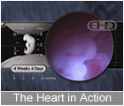
|
| Week 5 ends |
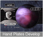
|
| Week 6 ends |
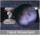
|
| Week 7 ends |
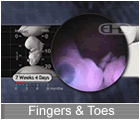
|
| Week 8 ends |
|
| Week 9 ends |
|
| Week 10 ends |
|
| Week 11 ends |
|
| Month 3 ends |
|
| Month 4 ends |
|
| Month 5 ends |
|
| Month 6 ends |
|
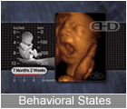
| Month 7 ends |
|
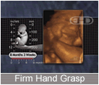
| Month 8 ends |
|
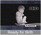
| Month 9 ends |
|
| Birth |
|
|
|
|
- (1 week, 1 day):
 Amnioblasts present; amnion and amniotic Amnioblasts present; amnion and amniotic- cavity formation begins
 Bilaminar embryonic disc Bilaminar embryonic disc Positive pregnancy test Positive pregnancy test-
- (1 week, 2 days): Carnegie Stage 5b
 Corpus luteum of pregnancy Corpus luteum of pregnancy Cells in womb engorged with nutrients Cells in womb engorged with nutrients Exocoelomic membrane Exocoelomic membrane Isolated trophoblastic lacunae Isolated trophoblastic lacunae Embryonic disc 0.1 mm diameter Embryonic disc 0.1 mm diameter-
- (1 week, 4 days): Carnegie Stage 5c
 Intercommunicating lacunae network Intercommunicating lacunae network Longitudinal axis Longitudinal axis Prechordal plate Prechordal plate Trophoblastic vascular circle Trophoblastic vascular circle- (1 week, 5 days):
 Implantation complete Implantation complete Yolk sac Yolk sac Embryonic disc diameter: 0.15 to 0.20 mm Embryonic disc diameter: 0.15 to 0.20 mm-
- (1 week, 6 days): Carnegie Stage 6
 Blood islands in umbilical vesicle Blood islands in umbilical vesicle Angiogenesis in chorionic mesoblast Angiogenesis in chorionic mesoblast Blood vessels in villi Blood vessels in villi Connecting stalk Connecting stalk Primordial blood vessels Primordial blood vessels Amnion with single cell layer Amnion with single cell layer Chorionic villi Chorionic villi- (2 weeks):
 Embryonic epiblast gives rise to Embryonic epiblast gives rise to- primitive streak and primitive node and
 Yolk sac Yolk sac Yolk sac Yolk sac- Less Events...
|
|
|
|
|
- (3 weeks, 1 day): Carnegie Stage 10
 Thyroid primordium emerges from floor of Thyroid primordium emerges from floor of- pharynx
 Nephrogenic cord emerges (at 10 somites) Nephrogenic cord emerges (at 10 somites) Cloaca Cloaca Common coelomic cavity divides into Common coelomic cavity divides into- peritoneal, pericardial, and pleural
- cavities
 Liver: Hepatic plate (endoderm) Liver: Hepatic plate (endoderm) Midgut emerging Midgut emerging Pharyngeal arches 1 and 2 Pharyngeal arches 1 and 2 Pharyngeal cleft 1 Pharyngeal cleft 1 Second pharyngeal cleft and pouch Second pharyngeal cleft and pouch Pharyngeal groove and ridge with Pharyngeal groove and ridge with- laryngotracheal sulcus
 Respiratory outgrowth Respiratory outgrowth Atria (right and left) far apart Atria (right and left) far apart Bulbis cordis Bulbis cordis Circulatory system function begins Circulatory system function begins Endocardial tubes fuse forming tubular Endocardial tubes fuse forming tubular- heart
 Heart begins beating Heart begins beating Pericardial sac Pericardial sac Pericardium Pericardium Primary head vein Primary head vein Sinus venosus Sinus venosus Tubular heart begins folding Tubular heart begins folding Umbilical arteries Umbilical arteries Umbilical veins (right and left) Umbilical veins (right and left) Optic primordia fill neuromere D2 Optic primordia fill neuromere D2 Chiasmatic plate Chiasmatic plate Mesencephalic flexure Mesencephalic flexure Neural tube Neural tube Neuromeres D1 and D2 (in diencephalon) Neuromeres D1 and D2 (in diencephalon) Optic sulcus in forebrain Optic sulcus in forebrain Pontine region identifiable near cranial Pontine region identifiable near cranial- nerves VII and VIII
 Segment D in rhombencephalon Segment D in rhombencephalon Some secondary neuromeres Some secondary neuromeres Superior colliculus Superior colliculus Telencephalon Telencephalon Telencephalon (or telencephalic) medium Telencephalon (or telencephalic) medium Body cavities Body cavities Hyoid arch Hyoid arch Mandibular arch and maxillary process Mandibular arch and maxillary process Neural crest: Trigeminal, facioacoustic, Neural crest: Trigeminal, facioacoustic,- glossopharyngeal-vagal, and
- occipitospinal
 Somites: Pairs 4 through 12 Somites: Pairs 4 through 12-
- (3 weeks, 3 days): Carnegie Stage 11
 Primordial germ cells begin moving from Primordial germ cells begin moving from- umbilical vesicle to hindgut
 Thyroid complete Thyroid complete Face: Maxillary and mandibular processes Face: Maxillary and mandibular processes- (bilaterally)
 Cloacal membrane Cloacal membrane Mesonephric duct emerges from Mesonephric duct emerges from- nephrogenic cord
 Nephric vesicles Nephric vesicles Cystic primordium Cystic primordium Hepatic diverticulum Hepatic diverticulum Liver Liver Membrane between future mouth and throat Membrane between future mouth and throat- may begin to rupture
 Angiogenesis along surface of central Angiogenesis along surface of central- nervous system
 Aortic sac Aortic sac Atrioventricular canal Atrioventricular canal Capillary plexus begins forming around Capillary plexus begins forming around- brain and spinal cord
 Conotruncus Conotruncus Conus cordis emerging from right Conus cordis emerging from right- ventricle
 Endocardium Endocardium Heart contractions produce peristaltic Heart contractions produce peristaltic- blood flow
 Internal carotid arteries Internal carotid arteries Interventricular septum Interventricular septum Primordium of myocardium Primordium of myocardium Sinus venosus separating from left atria Sinus venosus separating from left atria Trabeculated outpouches along primary Trabeculated outpouches along primary- cardiac tube representing primordia of
- left and right ventricles
 Trigeminal and otic arteries Trigeminal and otic arteries Facio-vestibulocochlear ganglia (CN VII, Facio-vestibulocochlear ganglia (CN VII,- CN VIII)
 Glossopharyngeal and vagal ganglia Glossopharyngeal and vagal ganglia Optic evagination (starting at 14 Optic evagination (starting at 14- somites)
 Otic vesicle Otic vesicle Trigeminal ganglia (CN V) Trigeminal ganglia (CN V) Adenohypophysial pouch Adenohypophysial pouch Adenohypophysis Adenohypophysis Lamina terminalis Lamina terminalis Mesencephalon contains tectum and Mesencephalon contains tectum and- tegmentum
 Neural crest production and migration Neural crest production and migration- continue
 Neurohypophysial primordia Neurohypophysial primordia Neuropore (near brain) closes Neuropore (near brain) closes Notochord Notochord Segmentation of mesoblast alongside Segmentation of mesoblast alongside- neural tube bilaterally
 Somites: Pairs 13 through 20 Somites: Pairs 13 through 20- (3 weeks, 3 days - 5 weeks, 6 days):
 All eight rhombomeres (Rh 1 through Rh All eight rhombomeres (Rh 1 through Rh- 7, Rh D) - Present in stages 11 through
- 17
-
- (3 weeks, 5 days): Carnegie Stage 12
 Telopharyngeal bodies Telopharyngeal bodies Alimentary epithelium invades stroma of Alimentary epithelium invades stroma of- liver
 Alimentary epthelium proliferates in Alimentary epthelium proliferates in- primordia of stomach, liver, and dorsal
- pancreas
 First part of pancreas First part of pancreas Gastric portion of foregut elongates (25 Gastric portion of foregut elongates (25- to 28 somites)
 Hepatic primordium with abundant Hepatic primordium with abundant- vascular plexus
 Omental bursa Omental bursa Oropharyngeal membrane is ruptured Oropharyngeal membrane is ruptured Pharyngeal arch 3 Pharyngeal arch 3 Pharyngeal arches with dorsal and Pharyngeal arches with dorsal and- ventral parts
 Umbilical vesicle elongates Umbilical vesicle elongates Cervical sinus Cervical sinus Laryngotracheal groove Laryngotracheal groove Lung bud Lung bud Tracheo-esophageal septum Tracheo-esophageal septum Atrioventricular canal Atrioventricular canal Common cardinal veins (right and left) Common cardinal veins (right and left) Descending aorta Descending aorta Heart circulates blood to and from Heart circulates blood to and from- central nervous system, umbilical
- vesicle, and chorion
 Hepatocardiac channels (right and left) Hepatocardiac channels (right and left) Rostral and caudal cardinal veins along Rostral and caudal cardinal veins along- brain and spinal cord feeding common
- cardinal veins
 Septum primum and foramen primum Septum primum and foramen primum- sometimes present
 Septum primum, foramen primum Septum primum, foramen primum Sinu-atrial foramen prevents backflow Sinu-atrial foramen prevents backflow- into sinus venosus
 Sinus venosus collects veinous blood Sinus venosus collects veinous blood- from entire embryo
 Superior vena cava, inferior vena cava, Superior vena cava, inferior vena cava,- and sinus venosus collecting all venous
- blood
 Unidirectional circulation Unidirectional circulation Vitelline arteries and veins Vitelline arteries and veins Hypoglossal cord (CN XII) enters Hypoglossal cord (CN XII) enters- pharyngeal arch 4
 Otocyst nearly closed Otocyst nearly closed Nasal discs form part of ectodermal ring Nasal discs form part of ectodermal ring Brain involves 40% of neural tube Brain involves 40% of neural tube Brain: Embryonic commissural plate Brain: Embryonic commissural plate Ectodermal ring complete Ectodermal ring complete Hypoglossal nucleus (CN XII) Hypoglossal nucleus (CN XII) Lowermost spinal cord formation begins Lowermost spinal cord formation begins Mamillary recess Mamillary recess Marginal layer in rhombencephalon Marginal layer in rhombencephalon Mesencephalic flexure at 90 degrees Mesencephalic flexure at 90 degrees Mesencephalon with two neuromeres: M1 Mesencephalon with two neuromeres: M1- and M2
 Motor neurons in basal plate of Motor neurons in basal plate of- rhombencephalon
 Neural tube closes (lower back) Neural tube closes (lower back) Neurofibrils form in rhombencephalon Neurofibrils form in rhombencephalon Primary neurulation ends Primary neurulation ends Primordia of ventral thalamus and Primordia of ventral thalamus and- subthalamus in diencephalon
 Sulcus limitans Sulcus limitans Sulcus limitans in midbrain Sulcus limitans in midbrain Somites: Pairs 21 through 29 Somites: Pairs 21 through 29 Upper limb primordium at level of Upper limb primordium at level of- somites 8 to 10
 Progressively C-shaped embryo Progressively C-shaped embryo-
- (4 weeks): Carnegie Stage 13
 Spleen primordia Spleen primordia Thymic primordia Thymic primordia Lower lip forms from merging of Lower lip forms from merging of- mandibular processes
 Melanoblasts in epidermis Melanoblasts in epidermis Skin is so thin, you can see through it! Skin is so thin, you can see through it! Gonadal ridge extends from C-7 to T-8 Gonadal ridge extends from C-7 to T-8- levels
 Primordial germ cells migrate to Primordial germ cells migrate to- mesonephric ridges
 Primordial germ cells number several Primordial germ cells number several- hundred
 Urorectal septum Urorectal septum Thyroid bilobed and attached to pharynx Thyroid bilobed and attached to pharynx- by thryoglossal duct
 Diaphragm primordia Diaphragm primordia Glomeruli emerge in mesonephros Glomeruli emerge in mesonephros Mesonephric duct attached to cloaca Mesonephric duct attached to cloaca Nephric tubules now S-shaped Nephric tubules now S-shaped Urogenital sinus Urogenital sinus Urorectal cleavage line Urorectal cleavage line Diverticulum ilei marks division between Diverticulum ilei marks division between- foregut and hindgut
 Esophagus primordia Esophagus primordia Intestines growing in length Intestines growing in length Mesentery from end of duodenum to Mesentery from end of duodenum to- proximal half of colon
 Opening between gut and umbilical Opening between gut and umbilical- vesicle decreases
 Pancreas: Ventral pancreas Pancreas: Ventral pancreas Pharyngeal pouches 1 through 4 Pharyngeal pouches 1 through 4 Pharynx Pharynx Pleuroperitoneal canals Pleuroperitoneal canals Small & large intestines Small & large intestines Stalk of umbilical vesicle lengthens and Stalk of umbilical vesicle lengthens and- narrows
 Stomach assumes shape of a spindle Stomach assumes shape of a spindle Umbilical vesicle at height of Umbilical vesicle at height of- development
 Vitelline duct Vitelline duct Bronchial buds Bronchial buds Lungs begin filling chest cavity Lungs begin filling chest cavity Mesenchyme from coelomic epithelium Mesenchyme from coelomic epithelium- surrounds esophagus and lung buds
 Trachea Trachea Anterior, middle, and posterior cerebral Anterior, middle, and posterior cerebral- plexuses
 Aorta branches include dorsal Aorta branches include dorsal- intersegmental, lateral segmental, and
- ventral segmental arteries
 Aortic arches 4 and 6 Aortic arches 4 and 6 Artery from the common iliac artery Artery from the common iliac artery- feeds each lower limb bud
 Atrioventricular bundle Atrioventricular bundle Cardiac contractions still under Cardiac contractions still under- myogenic control
 Celiac artery, superior and inferior Celiac artery, superior and inferior- mesenteric arteries
 Circulatory system "well established" Circulatory system "well established" Common iliac arteries (right and left, Common iliac arteries (right and left,- from dorsal aorta bifurcation)
 Contractions well coordinated and Contractions well coordinated and- sequential from sinus venosus to atria to
- ventricles
 Functioning two-chamber heart Functioning two-chamber heart Gas exchange through placenta begins Gas exchange through placenta begins Gelatinous reticulum (or cardiac Gelatinous reticulum (or cardiac- mesenchyme)
 Heart chambers bulging with fluid Heart chambers bulging with fluid Heart now functions as two parallel Heart now functions as two parallel- pumps
 Heart rate (about) 113 beats/min Heart rate (about) 113 beats/min Heart: Atrioventricular cushions Heart: Atrioventricular cushions- (rostroventral and caudodorsal)
 Heart: Myocardium wall 3 to 4 cells Heart: Myocardium wall 3 to 4 cells- thick
 Primary head veins (right and left) Primary head veins (right and left)- drain anterior, middle, and posterior
- cerebral plexuses and feed precardinal
- veins
 Small arteries emerging throughout Small arteries emerging throughout- mesoderm
 Ventricle walls trabeculated Ventricle walls trabeculated Vertebral arteries Vertebral arteries Vitelline veins empty exclusively into Vitelline veins empty exclusively into- hepatic plexus
 Most cranial nerve ganglia Most cranial nerve ganglia Trigeminal, glossopharyngeal, and vagal Trigeminal, glossopharyngeal, and vagal- preganglia
 Otic invagination Otic invagination Terminal-vomeronasal neural crest Terminal-vomeronasal neural crest Brain: Commissural plate Brain: Commissural plate Cerebellum Cerebellum Common afferent tract Common afferent tract Fourth ventricle Fourth ventricle Interstitial nucleus (part of medial Interstitial nucleus (part of medial- longitudinal fasciculus)
 Isthmus rhombencephali (a new neuromere) Isthmus rhombencephali (a new neuromere) Oculomotor (CN III) and trochlear nuclei Oculomotor (CN III) and trochlear nuclei- (CN IV) in mesencephalon (midbrain) and
- isthmus respectively
 Retinal and lens discs Retinal and lens discs Amnion surrounds connecting stalk and Amnion surrounds connecting stalk and- vitelline stalk
 Amnion surrounds embryo Amnion surrounds embryo Cervical flexure Cervical flexure Hyoid arch sudivides into dorsal and Hyoid arch sudivides into dorsal and- ventral segments
 Limb buds - the first sign of arms and Limb buds - the first sign of arms and- legs
 Lower limb buds Lower limb buds Umbilical cord emerging Umbilical cord emerging Upper and lower limb buds Upper and lower limb buds- Less Events...
|
|
|
|
|
- (5 weeks, 1 day):
 Wrist joints are forming Wrist joints are forming-
- (5 weeks, 2 days): Carnegie Stage 16
 Apical epidermal ridges Apical epidermal ridges Mammary ridge Mammary ridge Maxillary and premaxillary fields still Maxillary and premaxillary fields still- widely separated
 Nipples emerge from mammary crest Nipples emerge from mammary crest Gonad region separates from mesonephros Gonad region separates from mesonephros Gonadal primordium Gonadal primordium Labioscrotal swelling Labioscrotal swelling Urogenital fold and groove Urogenital fold and groove Suprarenal gland: Cortex primordium Suprarenal gland: Cortex primordium Suprarenal gland: Medulla Suprarenal gland: Medulla Thyroid detaches from pharynx Thyroid detaches from pharynx Thyroid with right and left lobes Thyroid with right and left lobes- connected by an isthmus
 Cartilage in mandibular arch Cartilage in mandibular arch Hand area with central carpal region and Hand area with central carpal region and- digital plate with marginal vein
 Pre-chondrocranium: Otic capsule, nasal Pre-chondrocranium: Otic capsule, nasal- capsule, and parachordal condensations
 Primordia of primary palate Primordia of primary palate Ribs: Primordia now present for all 12 Ribs: Primordia now present for all 12- pairs
 Vertebral column with 36 levels of Vertebral column with 36 levels of- ganglia and myotomes
 Extra-ocular premuscle masses receive Extra-ocular premuscle masses receive- cranial nerve fibers [oculomotor (CN
- III), trochlear (CN IV), and abducens (CN
- VI) nerves]
 Gluteal mesoderm Gluteal mesoderm Infrahyoid premuscle masses Infrahyoid premuscle masses Limb mesoderm Limb mesoderm Sternocleidomastoid-trapezius premuscle Sternocleidomastoid-trapezius premuscle- mass with spinal accessory nerve (CN11)
 Thigh and thigh mesoderm Thigh and thigh mesoderm Tongue premuscle mass Tongue premuscle mass Metanephros at level of sacrum Metanephros at level of sacrum Urethral plate Urethral plate Lesser omentum (ventral mesogastrun) Lesser omentum (ventral mesogastrun) Peritoneal cavity Peritoneal cavity Rectum Rectum Stomach: Greater and lesser curvatures Stomach: Greater and lesser curvatures Yolk stalk disappears Yolk stalk disappears Bronchial tree expanding Bronchial tree expanding Cervical sinus diminished in size Cervical sinus diminished in size Epiglottis Epiglottis Primitive Larynx Primitive Larynx Anterior, middle, and posterior cerebral Anterior, middle, and posterior cerebral- arteries
 Atrioventricular (AV) node Atrioventricular (AV) node Atrioventricular cushions fuse with Atrioventricular cushions fuse with- interventricular septum
 Circle of Willis almost complete Circle of Willis almost complete Conotruncal septum Conotruncal septum Endocardial cushions (rostroventral and Endocardial cushions (rostroventral and- caudodorsal) begin fusing around
- atrioventricular canal forming right and
- left atrioventricular canals and two
- separate blood streams
 External carotid artery External carotid artery Foramen primum disappearing Foramen primum disappearing Hepatic portal vein Hepatic portal vein Infundibulum of right ventricle Infundibulum of right ventricle Jugular lymph sac Jugular lymph sac Lateral atrioventricular cushions Lateral atrioventricular cushions Mesencephalic artery Mesencephalic artery Myelencephalic artery Myelencephalic artery Perilental blood vessels Perilental blood vessels Primitive cavernous sinus drains Primitive cavernous sinus drains- primitive maxillary and supraorbital
- veins
 Primitive renal plexus Primitive renal plexus Right ventricle feeds sixth (pulmonary) Right ventricle feeds sixth (pulmonary)- aortic arches; left ventricle feeds
- fourth aortic arches
 Semilunar valves (aortic and pulmonary) Semilunar valves (aortic and pulmonary)- are forming
 Ventricles each with three parts: inlet, Ventricles each with three parts: inlet,- trabecular pouch, and outflow tract
 Ventricles enlarge and deepen Ventricles enlarge and deepen- side-by-side forming an ever growing
- interventricular septum
 Celiac plexus Celiac plexus Cochlear nerve present Cochlear nerve present Femoral and obturator nerves innervate Femoral and obturator nerves innervate- rostrolateral part of lower limb
 Hypoglossal nerve (CN XII) reaches Hypoglossal nerve (CN XII) reaches- tongue
 Intercostal nerves Intercostal nerves Lumbar and sacral plexuses Lumbar and sacral plexuses Musculocutaneous, radial, ulna, and Musculocutaneous, radial, ulna, and- median nerves enter upper limb bud
 Nasal pits face more ventrally, still Nasal pits face more ventrally, still- widely separated
 Nasofrontal groove Nasofrontal groove Olfactory fibers connect nasal pits with Olfactory fibers connect nasal pits with- brain
 Olfactory fibers enter brain Olfactory fibers enter brain Olfactory tubercle present Olfactory tubercle present Peroneal and tibial nerves innervate Peroneal and tibial nerves innervate- caudomedial part of lower limb
 Phrenic nerve Phrenic nerve Pigment in retina visible externally Pigment in retina visible externally Primordium of cochlear pouch Primordium of cochlear pouch Tibial nerve innervates foot area Tibial nerve innervates foot area Auricular hillocks on hyoid arch Auricular hillocks on hyoid arch- (antitragus and helix)
 Auricular hillocks on mandibular arch Auricular hillocks on mandibular arch- (tragus and crus)
 Alar lamina emerging with dense rhombic Alar lamina emerging with dense rhombic- lip
 All cranial nerves identifiable All cranial nerves identifiable Archipallium, paleopallium, and Archipallium, paleopallium, and- neopallium
 Area epithelialis Area epithelialis Brain: Primordial plexiform layer in Brain: Primordial plexiform layer in- area of future temporal lobe
 Cajal-Retzius cells Cajal-Retzius cells Commissure of the trochlear nerve Commissure of the trochlear nerve Diencephalic subthalamic nucleus Diencephalic subthalamic nucleus Dorsal and ventral thalami separated by Dorsal and ventral thalami separated by- groove
 Dorsal funiculus fibers reach medulla Dorsal funiculus fibers reach medulla- oblongata
 Epiphysis cerebri Epiphysis cerebri Glial cells identifiable adjacent to Glial cells identifiable adjacent to- neurons
 Greater petrosal nerve Greater petrosal nerve Hippocampus: Gyrus dentatus Hippocampus: Gyrus dentatus Infundibular recess and infundibulum Infundibular recess and infundibulum Interventricular foramen large Interventricular foramen large Marginal ridge Marginal ridge Medial and lateral ridges of corpus Medial and lateral ridges of corpus- striatum are continuous
 Median forebrain bundle Median forebrain bundle Neurohypophysial outgrowth Neurohypophysial outgrowth Olfactory tubercle Olfactory tubercle Pontine flexure deepens Pontine flexure deepens Posterior commissure Posterior commissure Recurrent laryngeal nerve Recurrent laryngeal nerve Reticular formation more defined Reticular formation more defined Retinal fissure closes Retinal fissure closes Splanchnic nerve Splanchnic nerve Sulcus limitans hippocampi Sulcus limitans hippocampi Superior laryngeal nerve Superior laryngeal nerve Second pharyngeal arch more prominent Second pharyngeal arch more prominent Third pharyngeal arch recedes Third pharyngeal arch recedes- (5½ weeks):
 Initial tooth formation Initial tooth formation- (5½ weeks - 6 weeks):
 Subtle movement begins Subtle movement begins- (5 weeks, 4 days):
 Cartilage formation Cartilage formation- (5 weeks, 5 days):
 Nerve cells differentiating Nerve cells differentiating- (5 weeks, 5 days - 7 weeks, 1 day):
 Melanocytes in epidermis Melanocytes in epidermis-
- (5 weeks, 6 days): Carnegie Stage 17
 Facial growth centers grow and begin Facial growth centers grow and begin- merging forming nose and upper jaw
 Genital eminence forms phallus or Genital eminence forms phallus or- genital tubercle
 Gonad grows into oval shape with Gonad grows into oval shape with- irregular surface
 Auditory ossicles identifiable in Auditory ossicles identifiable in- mesenchyme
 Cartilage in occipital sclerotomes (1-4) Cartilage in occipital sclerotomes (1-4) Digital rays in hand plate Digital rays in hand plate Femur: Chondrification begins Femur: Chondrification begins Foot with rounded digital plate Foot with rounded digital plate Hypoglossal foramen (or canal) through Hypoglossal foramen (or canal) through- sclerotome 4 (area of future occipital
- bone)
 Odontogenic epithelium ermerges in six Odontogenic epithelium ermerges in six- areas (four maxillary and two mandibular)
 Primary palate components (right and Primary palate components (right and- left) fuse in midline
 Primitive palatine groove Primitive palatine groove Primordium of cartilage within nasal Primordium of cartilage within nasal- septum
 Vertebral centra begin chondrification Vertebral centra begin chondrification Primordia of orbital muscles Primordia of orbital muscles Calices Calices Mesonephros can produce urine Mesonephros can produce urine Pelvis of the ureter with three main Pelvis of the ureter with three main- divisions
 Vesico-urethral canal Vesico-urethral canal Biliary ducts within liver Biliary ducts within liver Dorsal and ventral pancreas fuse but Dorsal and ventral pancreas fuse but- retain separate ducts
 Duodenum enlarges proximal to and distal Duodenum enlarges proximal to and distal- to bile and pancreatic ducts
 Esophagus developing a submucous coat Esophagus developing a submucous coat- surrounding epithelium
 Intestinal loop begins umbilical Intestinal loop begins umbilical- herniation
 Primordial vermiform appendix Primordial vermiform appendix Stomach regions include gastric canal, Stomach regions include gastric canal,- fundus, corpus (or body), and pyloric
- antrum
 Trachea: Precursors of tracheal Trachea: Precursors of tracheal- cartilages
 Condensing mesenchyme around junction Condensing mesenchyme around junction- between left and right atria and cardiac
- tube is precursor to mitral and tricuspid
- valves
 Outflow tract rotates counterclockwise Outflow tract rotates counterclockwise Right and left atrioventricular canals Right and left atrioventricular canals- totally separated
 All parasympathetic cranial nerve All parasympathetic cranial nerve- ganglia identifiable
 All spinal nerves present All spinal nerves present Cell islands in olfactory tubercle Cell islands in olfactory tubercle Crescentic lens cavity Crescentic lens cavity Geniculate ganglion separate from Geniculate ganglion separate from- vestibulocochlear nerve
 Globular process emerges from each Globular process emerges from each- medial nasal process
 Nasal fin connecting nasal disc and Nasal fin connecting nasal disc and- surface epithelium
 Nasofrontal grooves Nasofrontal grooves Olfactory tubercle with cellular islands Olfactory tubercle with cellular islands Capillaries between adenohypophysis and Capillaries between adenohypophysis and- hypothalamus
 Commissure of the oculomotor nerves Commissure of the oculomotor nerves Cortical nucleus in amygdaloid body Cortical nucleus in amygdaloid body Dentate and isthmic nuclei in cerebellum Dentate and isthmic nuclei in cerebellum Dura begins forming in basal area Dura begins forming in basal area Epiphysis cerebri with intermediate Epiphysis cerebri with intermediate- layer
 First hint of septal nucleus First hint of septal nucleus Frontal and temporal poles of cerebral Frontal and temporal poles of cerebral- hemispheres
 Gustatory fibers separate from common Gustatory fibers separate from common- afferent tract
 Hemispheric stalk Hemispheric stalk Intermediate layer in tectum Intermediate layer in tectum- mesencephali
 Interventricular foramen Interventricular foramen Mesencephalon with intermediate layer Mesencephalon with intermediate layer Somites: Pairs 38 and 39 Somites: Pairs 38 and 39 Spinal cord reaches caudal tip of body Spinal cord reaches caudal tip of body Subarachnoid space Subarachnoid space Synapses in spinal cord between Synapses in spinal cord between- interneurons and primary afferent neurons
 Ventral thalamus with intermediate layer Ventral thalamus with intermediate layer- (6 weeks):
 Face withdraws from light touch around Face withdraws from light touch around- mouth
 Blood forming in liver Blood forming in liver Milk lines Milk lines Nipples along side of trunk Nipples along side of trunk Adrenal glands Adrenal glands Glucagon in pancreas Glucagon in pancreas Handplates develop subtle flattening Handplates develop subtle flattening Joints Joints Medial skull cartilages: Parachordal, Medial skull cartilages: Parachordal,- hypophyseal, and trabecular
 Tooth buds (primary teeth) Tooth buds (primary teeth) Diaphragm is largely formed Diaphragm is largely formed Intestines fill base of umbilical cord Intestines fill base of umbilical cord Synapses form in spinal cord Synapses form in spinal cord Crown-heel length 1.6 cm Crown-heel length 1.6 cm- Less Events...
|
|
|
|
|
- (7 weeks, 1 day): Carnegie Stage 20
 Facial processes no longer Facial processes no longer- distinguishable
 Ovaries full of primitive oogonia, Ovaries full of primitive oogonia,- intermediate pregranulosa cells, and
- mesenchyme
 Testes with short straight tubules Testes with short straight tubules Upper limbs with slightly flexed elbows Upper limbs with slightly flexed elbows Diaphragm: Central tendon Diaphragm: Central tendon Renal vesicles with S-shaped lumina Renal vesicles with S-shaped lumina Submandibular gland: Solid epithelial Submandibular gland: Solid epithelial- ducts enlarge and begin to branch
 Adenohypophysis with new capillaries on Adenohypophysis with new capillaries on- rostral surface
 Scalp vascular plexus Scalp vascular plexus Cochlear duct tip growing horizontally Cochlear duct tip growing horizontally Lens cavity completely filled Lens cavity completely filled Optic commissure Optic commissure Optic fibers extend to optic chiasma Optic fibers extend to optic chiasma Brain: Inferior colliculus (in Brain: Inferior colliculus (in- mesencephalon)
 Cerebral hemispheres expand beyond Cerebral hemispheres expand beyond- lamina terminalis
 Cerebral hemispheres extend over Cerebral hemispheres extend over- two-thirds of diencephalon
 Interpeduncular groove Interpeduncular groove Medial septal nucleus Medial septal nucleus Nigrostriatal fibers Nigrostriatal fibers Nucleus of diagonal band Nucleus of diagonal band Sacrocaudal spinal cord formation Sacrocaudal spinal cord formation- (secondary neurulation) complete
 Sensory pathways: Cuneate and gracile Sensory pathways: Cuneate and gracile- decussating fibers
 Septum verum Septum verum Spinothalamic tract Spinothalamic tract- (7 weeks, 1 day - 8 weeks):
 Stomach: Folds in stomach wall Stomach: Folds in stomach wall- (7 weeks, 2 days):
 Arteries and veins of heart complete Arteries and veins of heart complete-
- (7 weeks, 3 days): Carnegie Stage 21
 Volar pads begin to emerge on fingertips Volar pads begin to emerge on fingertips Chondrocranium with dorsum sellae and Chondrocranium with dorsum sellae and- hypophysial fossa
 Dens (of second cervical vertebrae) Dens (of second cervical vertebrae) Sternoclavicular joint and manubrium Sternoclavicular joint and manubrium The knee joints have arrived The knee joints have arrived Trachea: Thyroid cartilage Trachea: Thyroid cartilage Wrists slightly flexed Wrists slightly flexed Gluteus medius and gluteus minimus Gluteus medius and gluteus minimus- muscles
 Iliacus muscles Iliacus muscles Mylohyoid and infrahyoid muscles Mylohyoid and infrahyoid muscles Orbicularis oculi muscles Orbicularis oculi muscles Submandibular gland: Solid ducts with Submandibular gland: Solid ducts with- definitive branches
 Anterior and posterior choroid arteries Anterior and posterior choroid arteries Left superior vena cava disappears Left superior vena cava disappears- (Stages 21-23)
 Scalp vascular plexus moving toward Scalp vascular plexus moving toward- vertex
 Cornea: Substantia propria layer Cornea: Substantia propria layer Fibers of optic nerve reach brain Fibers of optic nerve reach brain Eyelids growing rapidly Eyelids growing rapidly Anterior and inferior horns of lateral Anterior and inferior horns of lateral- ventricle
 Brain: Insula within cerebral hemisphere Brain: Insula within cerebral hemisphere C-shaped lateral ventricle C-shaped lateral ventricle Cerebral hemispheres cover 75% of Cerebral hemispheres cover 75% of- diencephalon
 Cerebral hemispheres cover more than Cerebral hemispheres cover more than- half of diencephalon
 Cortical plate within primordial Cortical plate within primordial- plexiform layer
 Glial and neurilemmal (Schwann) cells Glial and neurilemmal (Schwann) cells- within cranial nerves
 Global pallidus internus Global pallidus internus Internal fiber layer of cerebellum Internal fiber layer of cerebellum Lateral olfactory tract Lateral olfactory tract Primordium of dentate nucleus Primordium of dentate nucleus Pyramidal cells in hippocampus Pyramidal cells in hippocampus Subthalamic nucleus proper, Subthalamic nucleus proper,- entopeduncular nucleus, and globus
- pallidus externus within subthalamus
 Sulcus transversus rhombencephali Sulcus transversus rhombencephali Ventral part of lateral geniculate body Ventral part of lateral geniculate body- (7½ weeks):
 Hands begin to touch face Hands begin to touch face The hands touch each other as do the The hands touch each other as do the- feet!
 Fingertips thicken Fingertips thicken Plantar pads toes Plantar pads toes EKG pattern similar to adult EKG pattern similar to adult- (7 weeks, 4 days):
 The fingers are free The fingers are free- (7 weeks, 5 days):
 Bone-forming cells called osteoblasts Bone-forming cells called osteoblasts- emerge
 Bone-forming cells emerge Bone-forming cells emerge Endolymphatic and jugular foramina Endolymphatic and jugular foramina Hands can reach one another and fingers Hands can reach one another and fingers- can overlap
 Optic foramen, foramen rotundum, Optic foramen, foramen rotundum,- internal acoustic foramen
 Osteoblasts emerge Osteoblasts emerge Pelvis: Obturator foramen Pelvis: Obturator foramen Obturator internus muscles Obturator internus muscles Rectus femoris muscle Rectus femoris muscle Large glomeruli present within Large glomeruli present within- metanephros
 Submandibular gland: Secondary branching Submandibular gland: Secondary branching- with lumen formation starting at oral end
- of duct
 Costodiaphragmatic recess of pleural Costodiaphragmatic recess of pleural- cavity
 Chordae tendineae (Stages 22 and 23) Chordae tendineae (Stages 22 and 23) Intradural veins (sinuses) Intradural veins (sinuses) Scalp vascular plexus 75% of the way to Scalp vascular plexus 75% of the way to- the vertex
 Cochlear duct's second loop growing Cochlear duct's second loop growing- upward
 Scleral condensation Scleral condensation Tragus and antitragus taking shape Tragus and antitragus taking shape Eyelids continue growing rapidly over Eyelids continue growing rapidly over- the surface of the cornea
 Brain: Claustrum Brain: Claustrum Brain: Cortical plate within cerebral Brain: Cortical plate within cerebral- hemispheres
 Brain: Internal capsule with connections Brain: Internal capsule with connections- to epithalamus, dorsal thalamus, and
- mesencephalon
 Brain: Putamen Brain: Putamen Cerebral hemispheres cover 75% of Cerebral hemispheres cover 75% of- diencephalon
 Commissural plate thickens Commissural plate thickens Cortical plate expanding rapidly Cortical plate expanding rapidly Folds in roof of third ventricle Folds in roof of third ventricle Nerve fibers between neopallial subplate Nerve fibers between neopallial subplate- and internal capsule
 Thalamocortical fibers Thalamocortical fibers- (7 weeks, 6 days):
 The toes are free The toes are free-
- (8 weeks): Carnegie Stage 23
 Complex response to touch Complex response to touch More frequent hand-to-face contact More frequent hand-to-face contact Mouth opens & closes Mouth opens & closes Squinting Squinting The embryo floats and rolls over in the The embryo floats and rolls over in the- womb
 Hairs first appear in eyebrows & around Hairs first appear in eyebrows & around- mouth
 Skin multi-layered, loses transparency Skin multi-layered, loses transparency Ductus deferens Ductus deferens Interstitial cells forming within testis Interstitial cells forming within testis Testicular tubules Testicular tubules Male embryos are making testosterone Male embryos are making testosterone- already!
 Anterior inferior iliac spine Anterior inferior iliac spine Costal cartilage Costal cartilage Enamel organ Enamel organ Femur: Head and acetabular fossa Femur: Head and acetabular fossa Glenoid fossa Glenoid fossa Greater trochanter Greater trochanter Head of humerus Head of humerus Inguinal ligament Inguinal ligament Joint development: Cavitation underway Joint development: Cavitation underway- in hip, knee, and ankle (in some embryos)
 Joint development: Cavitation underway Joint development: Cavitation underway- in shoulder, elbow, and wrist (in some
- embryos)
 Nucleus pulposus (from notochord) Nucleus pulposus (from notochord) Ossification underway in scapula and Ossification underway in scapula and- distal phalanges in some embryos
 Pubic symphysis Pubic symphysis Scapular spine and notch Scapular spine and notch Skull: Foramen magnum (wide) Skull: Foramen magnum (wide) Skull: Ossification underway in some Skull: Ossification underway in some- embryos
 Superior and inferior pubic rami Superior and inferior pubic rami The embryo's joints are similar to adult The embryo's joints are similar to adult- joints
 Ulna: Styloid process and olecranon Ulna: Styloid process and olecranon Vertebrae cartilaginous (33 or 34 in Vertebrae cartilaginous (33 or 34 in- number)
 Anterior digastric muscles Anterior digastric muscles Depressor anguli oris muscle Depressor anguli oris muscle Diaphragm complete Diaphragm complete Esophagus: Longitudinal muscles Esophagus: Longitudinal muscles Obliquus superior capitus muscle Obliquus superior capitus muscle Obturator externus, gluteus maximus, and Obturator externus, gluteus maximus, and- hamstring muscles
 Posterior belly of the digastric muscle Posterior belly of the digastric muscle Psoas tendon Psoas tendon Rectus sheath with anterior and Rectus sheath with anterior and- posterior lamina
 Temporal and lateral pterygoid muscles Temporal and lateral pterygoid muscles Zygomaticus major muscle Zygomaticus major muscle Kidneys at level of first three lumbar Kidneys at level of first three lumbar- vertebrae
 Metanephros: Numerous large glomeruli Metanephros: Numerous large glomeruli Metanephros: Secretory tubules Metanephros: Secretory tubules- elongating and becoming convoluted
 Sinusal tubercle Sinusal tubercle Urethra Urethra Urine production and release Urine production and release Gastrolienal ligament Gastrolienal ligament Nerves reaching intestinal loop Nerves reaching intestinal loop Peristalsis in large intestine Peristalsis in large intestine Submandibular gland: Lumen present in Submandibular gland: Lumen present in- terminal portions of duct
 Submandibular gland: Mesodermal sheath Submandibular gland: Mesodermal sheath- surrounds gland
 Unfused uvula (edge of unfused palatine Unfused uvula (edge of unfused palatine- shelf) and secondary palate
 Occasional breathing motions begin Occasional breathing motions begin Pseudoglandular stage begins Pseudoglandular stage begins Azygos vein Azygos vein Blood supply to the brain closely Blood supply to the brain closely- resembles adult pattern
 Hemiazygos veins Hemiazygos veins Inferior epigastric artery Inferior epigastric artery Inferior vena cava valve at junction of Inferior vena cava valve at junction of- right atrium
 Scalp vascular plexus nearing vertex Scalp vascular plexus nearing vertex Submandibular glands: Angiogenesis Submandibular glands: Angiogenesis- begins around epithelial tree (ducts)
 Superior sagittal sinus Superior sagittal sinus Cochlear duct's 2.5 coils nearly Cochlear duct's 2.5 coils nearly- complete
 Cranial nerve distribution mimics adult Cranial nerve distribution mimics adult- pattern
 Ear drum Ear drum Eye: Secondary vitrous body Eye: Secondary vitrous body Lens: Secondary lens fibers emerging Lens: Secondary lens fibers emerging Retina: Eight layers present Retina: Eight layers present Retina: Four of the ten adult layers Retina: Four of the ten adult layers- present
 Tympanic membrane Tympanic membrane Eyelids fusing laterally and medially Eyelids fusing laterally and medially Optic tract reaches ventral portion of Optic tract reaches ventral portion of- lateral geniculate body
 "The hindbrain "presents striking "The hindbrain "presents striking- resemblance to that of the newborn."
 "The rhombencephalon...presents striking "The rhombencephalon...presents striking- resemblance to that of the newborn."
 Amygdala area Amygdala area Brain represents 43% of embryo Brain represents 43% of embryo Brain: Caudate nucleus and putamen Brain: Caudate nucleus and putamen- within corpus striatum
 Cerebellar commissures Cerebellar commissures Cerebellum with external germinal layer Cerebellum with external germinal layer Cerebral hemispheres cover lateral Cerebral hemispheres cover lateral- portion of diencephalon
 Choroid plexus now lobular Choroid plexus now lobular Cortical plate covers nearly all of Cortical plate covers nearly all of- neopallial surface
 Dura lines entire vertebral canal Dura lines entire vertebral canal Fasciculus cuneatus and fasciculus Fasciculus cuneatus and fasciculus- gracilis form the decussation of the
- medial lemnisci
 Greater palatine nerve Greater palatine nerve Grey and white matter Grey and white matter Hippocampus reaches temporal pole Hippocampus reaches temporal pole Inferior and superior cerebellar Inferior and superior cerebellar- peduncles
 Most cisterns present Most cisterns present Principal nucleus of inferior olivary Principal nucleus of inferior olivary- nuclei
 Pyramidal decussations Pyramidal decussations Right- and left-handedness emerges Right- and left-handedness emerges Suprapineal recess Suprapineal recess Suprascapular nerve Suprascapular nerve Vermis of cerebellum Vermis of cerebellum Crown-heel length 4.3 cm Crown-heel length 4.3 cm Embryo contains approximately 1 billion Embryo contains approximately 1 billion- (10^9) cells
 Embryonic Period Ends Embryonic Period Ends The 8-week embryo has formed more than The 8-week embryo has formed more than- 4,000 permanent body parts.
- Less Events...
|
|
|
|
|
|
|
|
|
|
|
|
|
|
|
|
|
- (34 weeks):
 Rapid weight gain Rapid weight gain-
- (35 weeks):
 Firm grip Firm grip Amniotic fluid volume peaks Amniotic fluid volume peaks-
- (36 weeks):
 Surfactant production accelerates Surfactant production accelerates Brain weight 300 grams Brain weight 300 grams Crown-heel length 48.5 cm Crown-heel length 48.5 cm
|
|
|
|
|