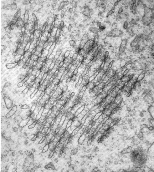
Very high power TEM of annulate lamellae in the cytoplasm of a stage 1b embryo in vitro, 24 hours post-insemination (original magnification x32,400). Shown are the pores, distended end of each lamella and intralamellar material of medium density which connects the edge of the pores in adjacent lamellae.
From: Soupart and Strong, 1974. Reproduced with permission of the American Society for Reproductive Medicine.
Keywords: annulate lamellae, cytoplasm, intralamellar material, lamella, stage 1b embryo
Source: The Virtual Human Embryo.