Your Life Before Birth – Read the Story
Introduction
You and everyone you know began your lives by successfully completing the journey before birth described in Your Life Before Birth. Read on to learn more about your journey and to better understand why the prenatal experience is so important for everyone.
When you share the movie and the following images as a simple act of friendship with those who have never seen similar images before, you enable them to join the growing numbers of people enlightened and enriched by the wonders, the beauty, and the significance of every pregnancy.
I began this work in a spirit of something like active curiosity, I have prosecuted it with an ever-deepening interest, and I have brought it thus far with the growing sense that I have been dealing with a subject of tremendous importance for the future of the [human] race and the individual....
J. W. Ballantyne, M.D.
(The "Father of Prenatal Care")
Manual of Antenatal Pathology and Hygiene - The Foetus. 1902
FROM THE SCRIPT: “Welcome to the amazing world inside the womb where every tiny baby grows and prepares for life after birth. It's also the place where bonding between mother and child begins and where each little one's lifelong health is partly established.”
A very busy time and place
A word about prenatal age
All prenatal ages on this page are referenced from the start of fertilization, not the beginning of a woman's last menstrual period (LMP). To calculate the equivalent prenatal age referenced from a woman's LMP, add two weeks to the fertilization ages provided here.
A time of preparation
The nine months of pregnancy truly is a time of preparation for life after birth. With rare exception, virtually everything that a newborn baby can do has been practiced over and over again for weeks or months before birth.
A bond like no other
From the time of fertilization (or conception), the developing baby and mother seamlessly work together and communicate with one another in numerous and complex ways that are still not fully understood. Pregnancy is the ultimate expression of selfless teamwork as mother and child do everything, share everything, and experience everything together as they jointly pursue a healthy full-term pregnancy and safe delivery.
As you might expect, a mom’s health and well-being are closely intertwined with the health and well-being of her child. With few exceptions, what is good for mom is good for her baby, and what is good for her baby is good for mom. Likewise, what is harmful to mom is harmful to her baby, and what is harmful to her baby is harmful to mom.
The exceptional teamwork between mother and child begins almost immediately. The first pregnancy hormone, called early pregnancy factor, appears in mom's blood as early as 24 hours after fertilization. This hormone starts preparing her body to meet her baby’s needs.1
Prenatal growth and development influence lifelong health
Research over the past 30 years has uncovered many ways in which a baby’s prenatal environment and growth pattern influence life-long health.
…good paths of growth and development are the greatest gift society can give to the next generation.
David J. P. Barker, M.D.
Nutrition in the Womb: How better nutrition during development will prevent heart disease, diabetes, and stroke. 2008. Page 165.
To cite just a few examples, babies experiencing impaired prenatal growth face an increased risk of heart attack,2 stroke,3 hypertension,4 and Type II diabetes5 later in life. Tobacco exposure during pregnancy is associated with an increased risk of certain behavior disorders6 as well as obesity,7 diabetes,8 and diminished lung function.9
…It is evident that the fetal environment is of tremendous importance during the developmental period in determining health throughout the life of the individual.
Dr. Hugo T. Bergen
Associate Professor
Department of Anatomy and Cell Science
University of Manitoba
Fertilization – The Beginning of a New Individual
FROM THE SCRIPT: “ 'Human development begins at fertilization10' when a male and a female reproductive cell join to form a unique, single-cell embryo.”11
Fertilization involves a series of events, which lasts about 24 hours (rather than a moment) in humans.12 Fertilization typically occurs within a woman’s uterine tube, which extends from a woman’s ovary on each side, to her uterus or womb.
Human fertilization
Single-cell embryo
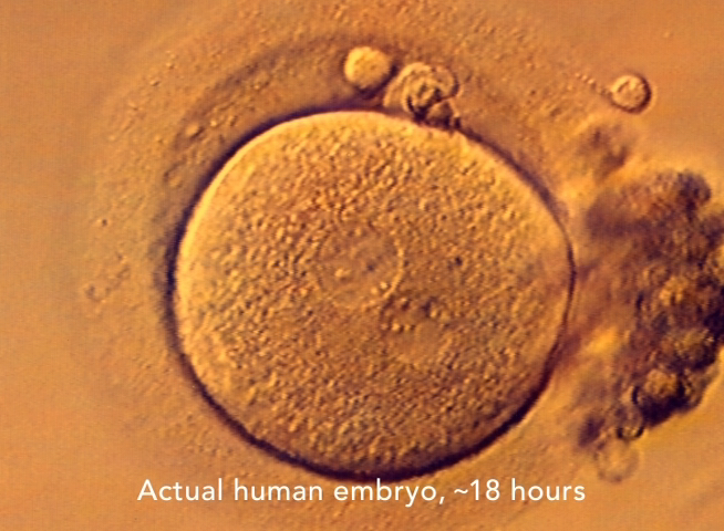
Human development and human life begin simultaneously
There are a number of unassailable scientific facts that you should know about human fertilization and the single-cell human embryo. All are important. Despite unequivocal scientific evidence, some remain controversial.
Fertilization, or conception, marks the beginning of human development; the start of a brand new, unique, genetically distinct human being; and the true onset of pregnancy.
The living, single-cell human embryo formed at fertilization is a new human life at the first stage of human development.13
Embryonic life commences with fertilization...
Ronan O'Rahilly and Fabiola Müller
Developmental Stages of Human Embryos
Carnegie Institute of Washington, 1987, Page 9.
It is to be remembered that at all stages the embryo is a living organism, that is, it is a going concern with adequate mechanisms for its maintenance as of that time.
C. H. Heuser and G. L. Streeter, 1941
Contributions to Embryology, No 181
Vol 29, No 181. Pages 15–55.
Because of the controversy, this topic deserves some further discussion.
Defining the start of human life as occurring at some point later than fertilization ignores and contradicts longstanding scientific fact. This is very simply understood. Only a continuously living embryo can divide from one cell into two, two cells into four cells, and so on throughout pregnancy. It is equally true that a dead embryo cannot grow, cannot divide, cannot change, cannot consume oxygen, cannot produce carbon dioxide, cannot replicate DNA, and cannot become alive at some future time.
Stage 1 is the unicellular embryo.... It is the beginning of embryonic life...
Raymond Gasser, Ph.D.
Principal Investigator, The Virtual Human Embryo Project
2001–2013. Stage 1. Introduction.
As a scientific question, to argue that the human embryo or fetus comes alive at some point beyond fertilization is to support a modern version of the theory of spontaneous generation. Proponents of this once-popular theory believed that living things could form from dead matter. This theory was debunked 160 years ago.
The distinction about when human life and human development begin is more than an academic or theoretical question. It has health implications. Once fertilization is underway, the fragile early embryo is susceptible to damage from the surrounding environment, which includes exposure to toxins present in mom’s uterine tube or intra-uterine environment.14
An amazing skill set
In addition to the ability to work closely with mom, the simple-appearing, single-cell human embryo directs its own development throughout life using the instruction set present in its own DNA.15
Comprehend if you can, how the most minute fragment of life—a single cell, just on the fringe of the microscopic world—can divide and redivide to make the billions and billions of cells in the body of a baby. Think of the uncountable number of tissues and organs that sprang from this initial fleck of clay: the hair, the nails, the skin, the brain, the eyes, the ductless glands. Each step of the creative process represents a series of exquisitely intricate events, each timed to the other in hairline sequence.
Better Homes and Gardens Baby Book—A Child Care and Training Guide
Meredith Publishing Company
1956. Page 15.
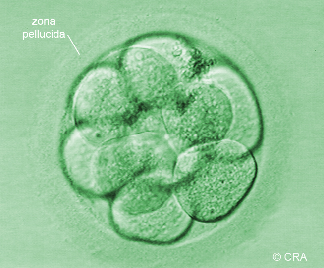
Multiplying and Dividing
FROM THE SCRIPT: “At about 24 hours, the single-cell embryo divides into two cells, then four cells, and so on. This begins the incredible transformation from single-cell embryo to precious newborn.”
Human embryo time-lapse (1–8 cells)
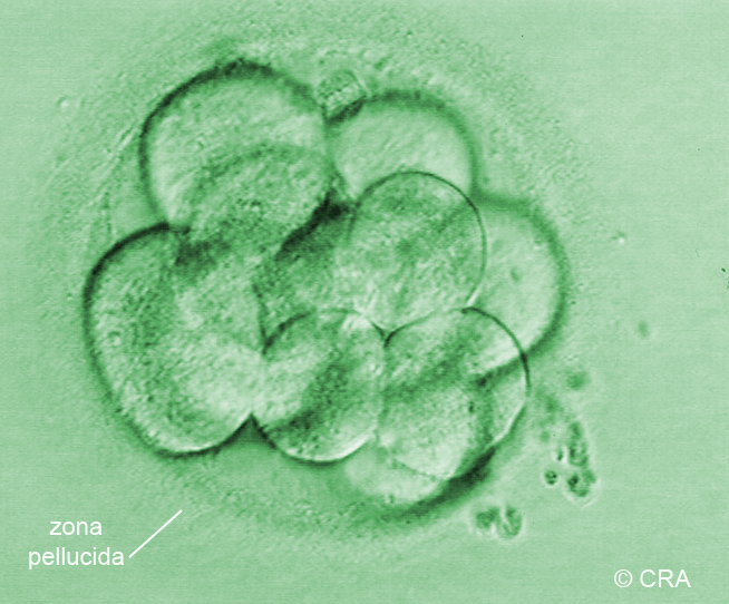
Cell division continues. The embryo reaches 12–16 cells after about three days and is temporarily shaped like a ball of cells. At this stage, the embryo is called a morula.16
10-cell human embryo
Human morula
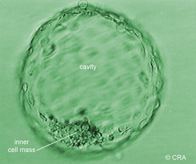
As cells continue to divide, a fluid-filled cavity emerges and the embryo is now called a blastocyst.
Early human blastocyst
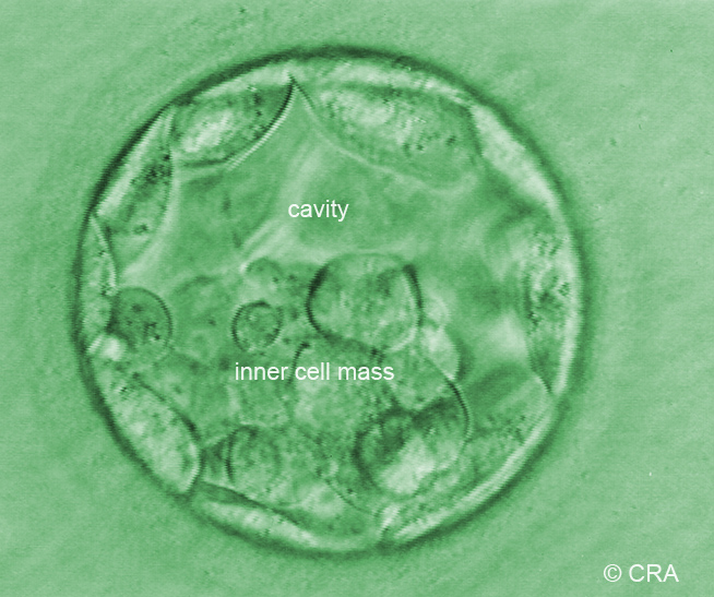
Mature human blastocyst
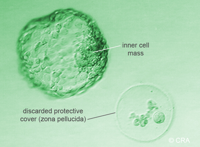
For normal development to proceed, the blastocyst must escape its protective coating. When viewed under the microscope, this process is called "hatching." The protective coating of the human embryo developing naturally inside the womb is believed to break down and fall apart, rather than "hatch" as seen here.
Early hatching blastocyst
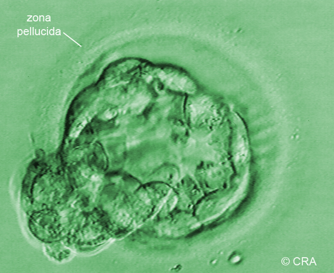
The great escape
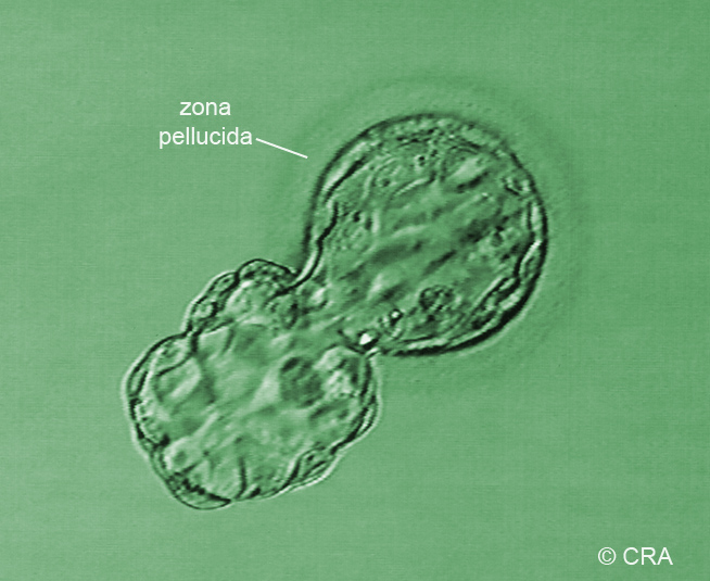
Once free, the embryo is ready for the next big step: implantation.
Free blastocyst
Implantation is the process whereby the embryo leaves the cavity of the uterus by embedding itself into the wall of the uterus. This process begins on day 6 with attachment to the uterine wall and ends roughly 12 days following fertilization.
The speed and complexity with which human development unfolds may surprise you. With repeated cell divisions and various specialized cell types forming along the way, the tiny baby grows and repeatedly changes shape. Human development progresses rapidly in a highly organized fashion.
The first sign of brain development appears at 18 days.17
The mere existence of that cell [referring to the cell giving rise to the human brain] should be one of the greatest accomplishments of the earth. People ought to be walking around all day, all through their waking hours, calling to each other in endless wonderment, talking of nothing except that cell... If anyone does succeed in explaining it, within my lifetime, I will charter a skywriting airplane, maybe a whole fleet of them, and send them aloft to write one great exclamation point after another, around the whole sky, until all my money runs out!
Lewis Thomas
The Medusa and the Snail, 1979
The basic body plan featuring distinct body regions such as the head, chest region, abdomen, and the beginnings of upper and lower limbs is well established by just 4 weeks after fertilization.18
Key Periods of Prenatal Development
FROM THE SCRIPT: “You may be surprised to know that most body parts form and begin to function in the first 8 weeks, even though full-term pregnancy lasts 38 weeks.”
Human development before birth is a front-loaded process. The first 8 weeks features rapid changes in shape and appearance, along with a dramatic rise in the number of identifiable body parts. Thereafter, human development is largely marked by growth and maturation of existing body parts and functions.
Therefore, human development before birth is divided into two periods.
The embryonic period begins with fertilization and ends following the completion of 8 weeks or 56 days.19 Throughout this entire time, the developing human is properly called an embryo, meaning “growing within.”20 Most body parts and all body systems first appear during this time and begin to function.21
The fetal period begins just as soon as the embryonic period ends and continues until birth. During this time, the developing human is called a fetus, meaning “unborn offspring.”22 During the fetal period, the baby grows much larger and body functions grow more and more complex.23
The Rapidly Developing Beating Heart
FROM THE SCRIPT: “By 22 days, the heart begins to beat... and quickly resembles the heart of a newborn.”
The first sign of heart development occurs at about 20 days.24 By 22 days, the tube-shaped heart begins to beat.25Almost immediately, the heart begins changing shape as seen below.26 As we will see time and time again, the developing human begins performing new functions at what seems to be the earliest possible opportunity.
The rapidly maturing heart
This early onset of the baby’s heartbeat has been reported at least as early as 1962 and is not theoretical.27 Researchers have reported visualizing the subtle contractions of the tiny human beating heart using ultrasound at 23 days28 and 25 days29 after fertilization. Although once believed to be true, there is no credible modern evidence to support the onset of the beating heart at 18 days or earlier, as some have proposed.
FROM THE SCRIPT: “By 4 weeks, the baby is surrounded by a fluid-filled sac, and her heart is pumping nutrients to her entire body.”
The fluid-filled sac is a tough membrane called the amnion. The fluid within the sac is called amniotic fluid.30 Together, they protect the growing baby from trauma, help maintain fluid balances during the first half of pregnancy, and provide room for the baby to move relatively free from the force of gravity.
The circulation
FROM THE SCRIPT: “Here you can see her beating heart in slow motion just 4½ weeks after fertilization. Do you see how her heart changes color as blood enters and leaves its chambers with each beat?”
The heart begins to beat at just about the earliest possible time that it could beat. By 4 weeks, blood circulated by this tiniest of all hearts is delivering life-sustaining oxygen and nutrients to various parts of the brain and body.31 Note the precision with which this little heart does its vital work!
The brain and beating heart
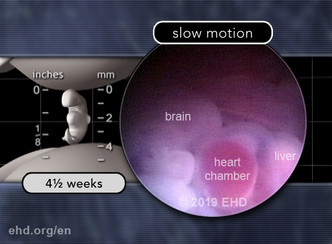
Beating heart in slow motion
The four-chambered beating heart
The circulatory system faces the daunting task of having to grow and become continuously remodeled to keep pace with the embryo's overall growth while remaining fully functional in supplying the needs of the embryo's cells.
Bruce M. Carlson, M.D., Ph.D.
Human Embryology and Developmental Biology
Third edition, Philadelphia: Mosby
2004. Page 114.
Movement and Brainwaves
FROM THE SCRIPT: “By 6 weeks, the baby begins to move and will turn away if lightly touched on his face. Brainwaves have been recorded as early as 6 weeks and 2 days.”
Movement begins
Movement begins just 5½ to 6 weeks after fertilization.32 It is one of our earliest activities and one of the most natural things we can do.
The baby's ability to sense and respond to a light touch on the face represents the first evidence of a working reflex pathway.33
You may be surprised to know that movement before birth is necessary for normal development of the nerves, bones, and joints. In fact, without movement early in pregnancy, normal joint formation would be impossible.34 Movement is vital to human health, starting long before birth and at all ages thereafter.35
Exploring new features
The earliest recorded brainwaves
The earliest report of brainwaves was obtained from direct measurements from an embryo estimated to be 44 days postfertilization (or 6 weeks and 2 days).36
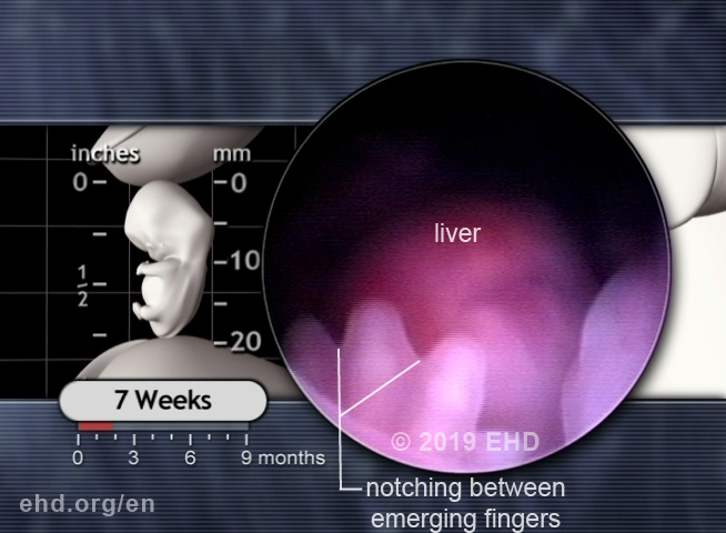
The 7-Week Embryo
FROM THE SCRIPT: “By 7 weeks, the baby begins turning her head and moving her hands.”
Actively engaged
Once movement begins, it doesn’t take long for many other movements of increasing complexity to emerge. These movements include coordinated muscle activity inside the body. Peristalsis is the complex traveling wave of coordinated muscle relaxation and contraction of the intestinal wall, which propels swallowed nutrients through the digestive tract. Peristalsis begins in the large intestine at 8 weeks37 and in the small intestine at 9 weeks.38
Fingers and Toes, Amazing Eyes
FROM THE SCRIPT: “Individual fingers are emerging from his hand plates… and his eyes are developing rapidly.”
The origin of fingers and toes
Distinct fingers and toes emerge from the hand and foot plates over about 10 or 11 days, starting at about 6 weeks after fertilization. It is well established that the cells between the individual fingers and toes, having served their purpose, will self-destruct through a process of planned (programmed) cell death.39
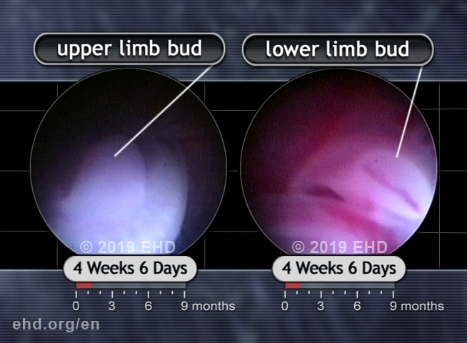
Upper and lower limb buds
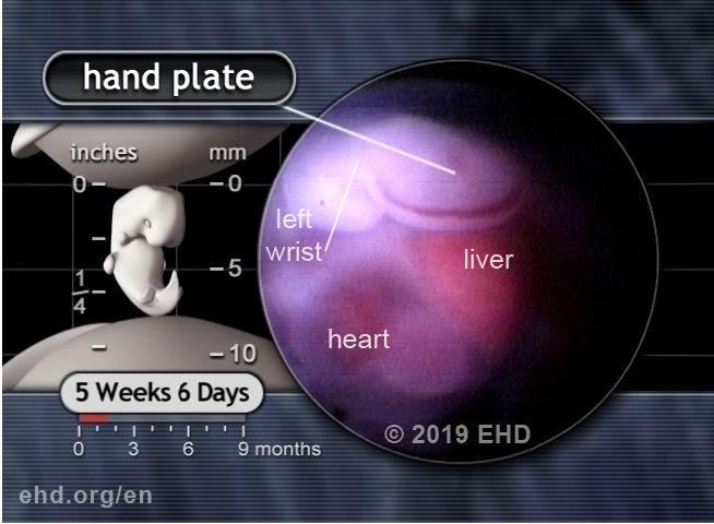
Hand plate and wrist
Now, even more dramatic changes are about to unfold.
Hand plate
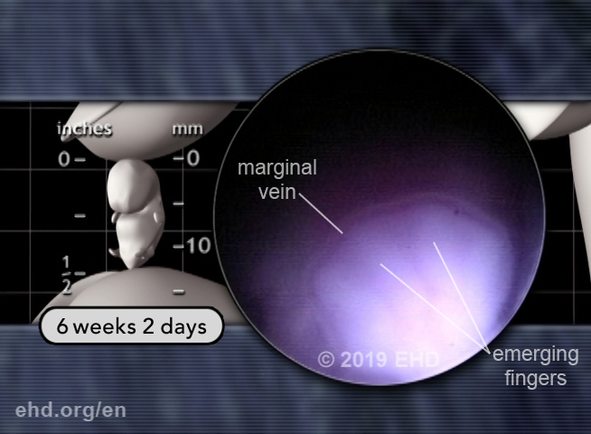
Emerging fingers
Fingers
Finger movement
The development of individual toes mimics that of the fingers but occurs several days later.
Eyes
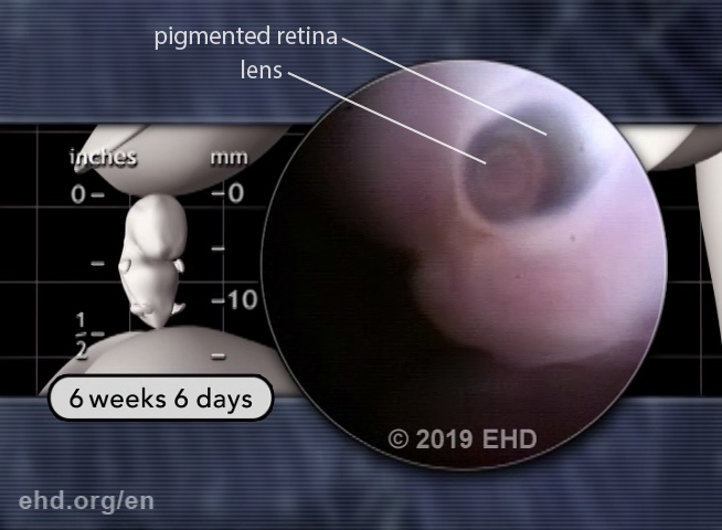
The eyes at 7 weeks are well formed but still not complete. The optic nerves are forming behind each eye, along with several layers of the retina.40 Several muscles that attach to and move each eye are emerging as well.
The maturing lens
FROM THE SCRIPT: “Leg movements are also seen... along with the startle response.”41
Legs and genital tubercle
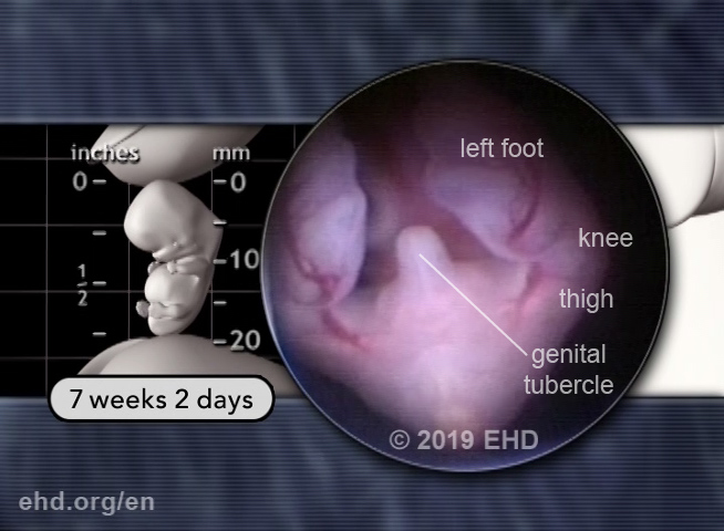
Leg movement, startle response
FROM THE SCRIPT: “The baby’s four-chambered heart is now nearly complete42 and continues its life-sustaining activity, resting only briefly between each heartbeat.”
Heartbeat
The human heart beats approximately 54 million times before birth.43 And that is just the beginning, as EHD further estimates that the human heart beats in excess of 3.2 billion times over a typical 80-year lifespan.44
Investigators have recorded an electrical tracing of the human heart at 7½ weeks following fertilization. By this age, the conducting system within the heart is very well developed, and the EKG tracing has a very similar appearance to EKG tracings obtained from newborns and even adults.45
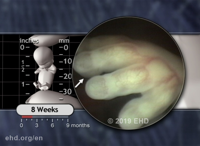
FROM THE SCRIPT: “By 7½ weeks, the baby’s fingers are separate. His hands begin to touch in the midline... as do his feet.”46
As his hand plates grow, the cells between the emerging fingers (and toes) undergo planned (programmed) cell death to create separation between the fingers.47 The baby’s hands can now reach and touch one another in the midline (with the feet doing likewise). This marks the beginning of exploring his world through the touch of a finger.
FROM THE SCRIPT: “Here you can see her beautiful right eye surrounded by her emerging eyelids.”
The human eye at this stage shows considerable similarity to the newborn and adult eye. Here you can see the developing lens and anterior chamber, behind which the relatively large posterior chamber is seen.
Eyelids
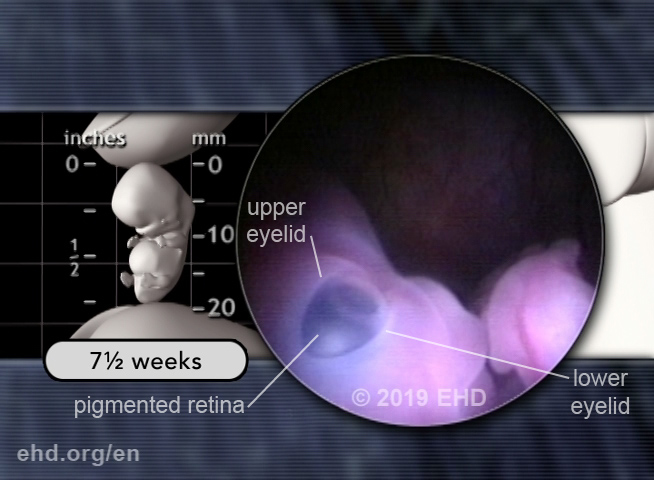
Within the posterior chamber of the eye, you can see the dark pigment of the human retina. Though not yet in final form, it has already been in place for weeks!48 You can also clearly see the early upper and lower eyelids. At this point, the eyelids begin to rapidly cover the entire surface of the eye and will soon fuse together.
The Amazing 8-Week Embryo
FROM THE SCRIPT: “Between 7 and 8 weeks, the baby forms nearly 2,000 additional body parts.”49
The sheer number of newly identifiable permanent body parts that emerge during the 7-day period from 7 weeks (or 49 days) to 8 weeks (56 days) is astounding. Despite the baby’s small size and young age, a whole new level of exquisite detail emerges as numerous individual ligaments, tendons, muscles, nerves, and blood vessels become identifiable for the first time.
The arteries of the hand
The arteries that supply the hand provide an excellent example of the exquisite detail present in the 8-week embryo. This screen shot captures the tiny arteries that supply the hand and fingers.
Hand arteries
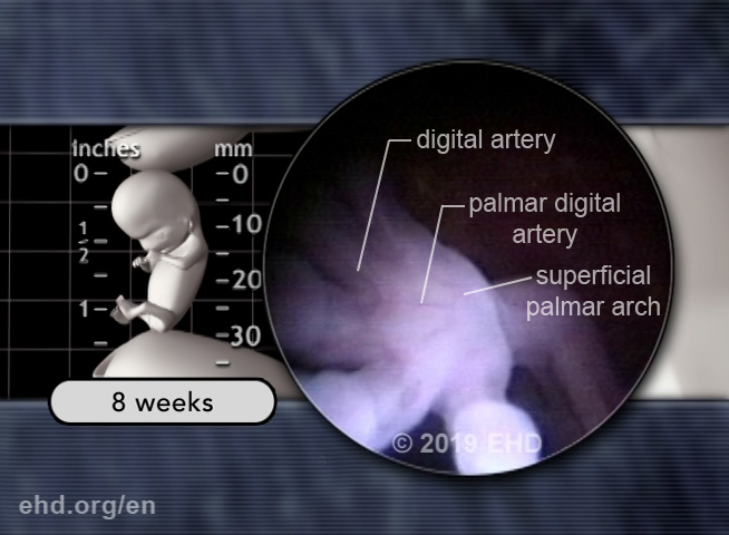
Hand arteries
By the end of the embryonic proper (8 postovulatory weeks), all of the major skeletal, articular, muscular, neural, and vascular elements of the limbs are present in a form and arrangement closely resembling those of the adult.
O'Rahilly R and Gardner E.
The timing and sequence of events in the development of the limbs in the human embryo.
Anat Embryol. 148(1):1-23.
1975. Page 15.
8-week embryo details
The 8-week human brain
FROM THE SCRIPT: “By 8 weeks, the baby’s brain is so complex that parts of it closely resemble the brain of a newborn.”50
Parts of the brainstem (the base of the brain closest to the spinal cord) and the arterial blood supply of the brain appear very similar to those found in the newborn.
Apart from differences in proportion, the arterial pattern resembles closely that of the adult. [Referring to the arterial blood supply of the human brain 8 weeks after fertilization]
Ronan O'Rahilly and Fabiola Müller
The embryonic human brain - An atlas of developmental stages.
Second edition. New York: Wiley-Liss.
1999. Page 332.
The arrangement of the rhombencephalic nuclei and tracts at 8 weeks is very similar to that present in the newborn.
Ronan O'Rahilly and Fabiola Müller
The embryonic human brain - An atlas of developmental stages.
Third edition. New York: Wiley-Liss.
2006. Page 219.
Because the human brain is so advanced by 8 weeks, some experts believe brain function is likely already underway.
The brain at stage 23 [8 weeks postfertilization] is far more advanced morphologically than is generally appreciated, to such an extent that functional considerations are imperative....It is likely that the rapid growth of the rhombencephalon during the embryonic period proper is associated with correspondingly early functional activity.
Ronan O'Rahilly and Fabiola Müller
The embryonic human brain - An atlas of developmental stages.
Third edition. New York: Wiley-Liss.
2006. Page 219.
FROM THE SCRIPT: “The baby begins breathing movements even though there is no air in the womb. Even at this early age, most babies prefer to use their right or left hand, just like you do.”
Preparing for first breath and first cry
Breathing movements before birth help blood return to the heart and prepare the breathing muscles for their vital duties after birth. At 8 weeks, breathing motions are observed only 2% of the time.51 As the baby matures, these motions are seen roughly 30%–40% of the time.52
A full-term newborn baby has the ability to draw in her first deep breath and release her first cry because she has prepared for this moment for 30 weeks! Her many hours spent practicing breathing motions has also given her the endurance to continue breathing.
Right- and left-handedness
Ultrasound studies have shown that 75% of babies demonstrate a definite preference to move their right hands, starting 8 weeks after fertilization. The remaining babies are split into two equal groups, with one group preferring to move their left hands and the other group showing no preference.53
Eyelids
Appreciating what has been accomplished
FROM THE SCRIPT: “In just 8 weeks, the remarkable single-cell embryo has blossomed into one billion cells, which have already formed an estimated 4,000 body parts54 and a dozen body systems.”
The astonishing transformation in just 8 weeks is incomprehensibly complex and, considering the incalculable number of steps involved, amazingly error free.
Just imagine the excitement when you find out how many structures you can recognize in a 5-week-old embryo, barely measuring 1 cm in length. But our fascination does not stop here. The progression of changes taking place in the next 3 weeks is so rapid that at the time when the embryo measures 3 cm, all structures familiar to us are not only easily recognizable, but also already in their anatomical position. How can we hide our amazement when we realize that such a state of perfection is present in an embryo a bit longer than the distal phalanx of our little finger?
Professor Hans K. Uhthoff, M.D., FRCS(C)
The embryology of the human locomotor system
Berlin: Springer-Verlag
1990. Preface.
One immediate consequence of this dramatic developmental progress is that now more than ever, the overall form of the 8-week human embryo closely resembles the overall shape of a newborn baby despite still being a tiny fraction of a newborn’s size.
This undeniable similarity is not coincidental. Rather, it is the inevitable consequence of the fact that the 8-week embryo and the newborn baby each possess thousands of the exact same body parts, and those parts are arranged in almost exactly the same way. With these facts in mind, a similar external appearance should be expected.
The Fetal Period
The fetal period begins 8 weeks and one day after fertilization and continues until birth.
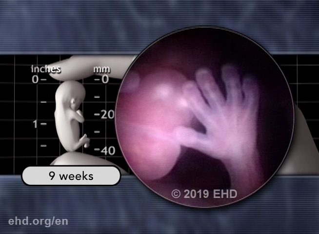
FROM THE SCRIPT: “By 9 weeks, the baby begins to suck his thumb,55 swallow,56 sigh,57 and stretch.58 His face, hands, and feet now sense and respond to light touch.”59
Active motion
The early fetus continues to respond to her environment in new ways. Swallowing is a complex function that is added to the growing list of abilities being practiced before birth.
With nearly every major nerve pathway in place between the skin and the spinal cord and even to parts of the brain, the baby is already extraordinarily advanced and enters a period of rapid growth.
FROM THE SCRIPT: “Yawning begins by 9½ weeks.60 By 10 weeks, he practices walking motions and his fingerprints begin to form.”
Yawning
A yawn is no simple thing. It involves an unusually deep inspiration with slight lifting of the head, followed by a deep expiration.
Fancy footwork
The fancy footwork you see here is, of course, just the beginning. Coordinated walking motions begin long before birth, but they become more difficult to practice as the baby grows, as room inside the womb becomes extremely limited before birth.
People dance at any age.
Mikhail Baryshnikov
The beginning of dance
Fingerprints
Fingerprints are unique identifiers and they are forming by 10 weeks.61
Fingernails and toenails are also beginning to grow.62
FROM THE SCRIPT: “By 11 weeks, the baby's mouth and lips are fully formed63 and she continues to grow much larger.”
There are approximately 30 muscles involved with facial expression.64 By 11 weeks, the baby begins to coordinate many of these muscles to make various expressions.65
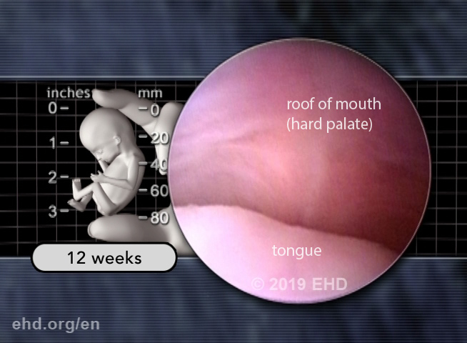
FROM THE SCRIPT: “By 12 weeks, the baby opens and closes her mouth, and moves her tongue. Her hands are fully formed.”
In the following screen captures, we can see the tongue and roof of the mouth. Look familiar? Their appearance already very much resembles those seen in newborns.
Roof of the mouth (hard palate)
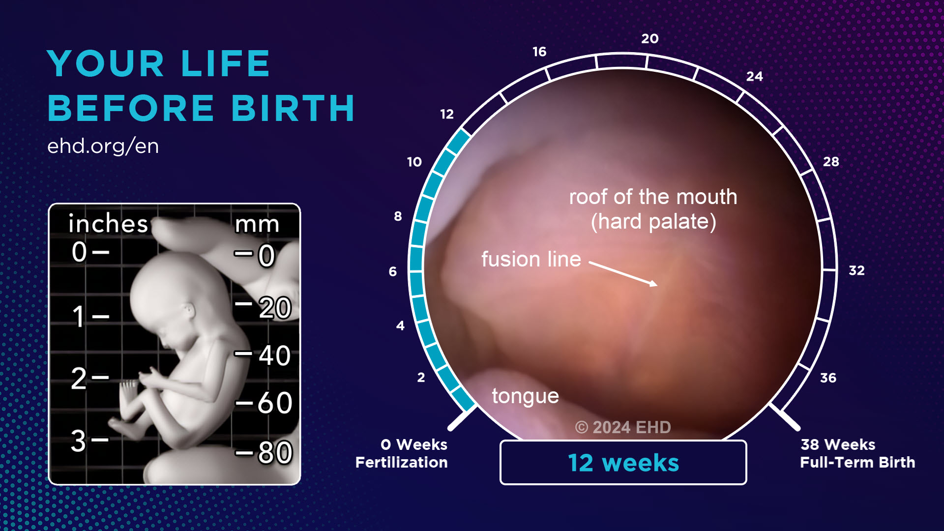
The tongue and roof of the mouth
The nervous system continues to mature. By 13 weeks, her ability to detect a light touch extends everywhere except parts of her scalp.66
She now begins to store fat.67 The emergence of fat deposits helps the baby to fill out. Fat represents stored energy and helps the baby regulate body temperature after birth.
FROM THE SCRIPT: “As pregnancy continues, the baby continues to grow and acquire new skills. Hearing,68 sleep-wake cycles,69 responding to sound70 and light,71 forming taste preferences,72 recognizing mom’s voice,73 and more, are all a routine part of the fascinating prenatal journey.”
It was not that many years ago that doctors believed smiling did not begin until 6 weeks after birth.74 We now know that smiling, like so many other typical human behaviors, is well rehearsed, starting long before birth.
FROM THE SCRIPT: “Pregnancy is indeed a time of preparation for life after birth. It is also a time to celebrate and anticipate the joys, wonders, and challenges that come with every new life.”
Newborn Baby
FROM THE SCRIPT: “Please share this free video with your family and friends. You can download it now at: ehd.org/en.”
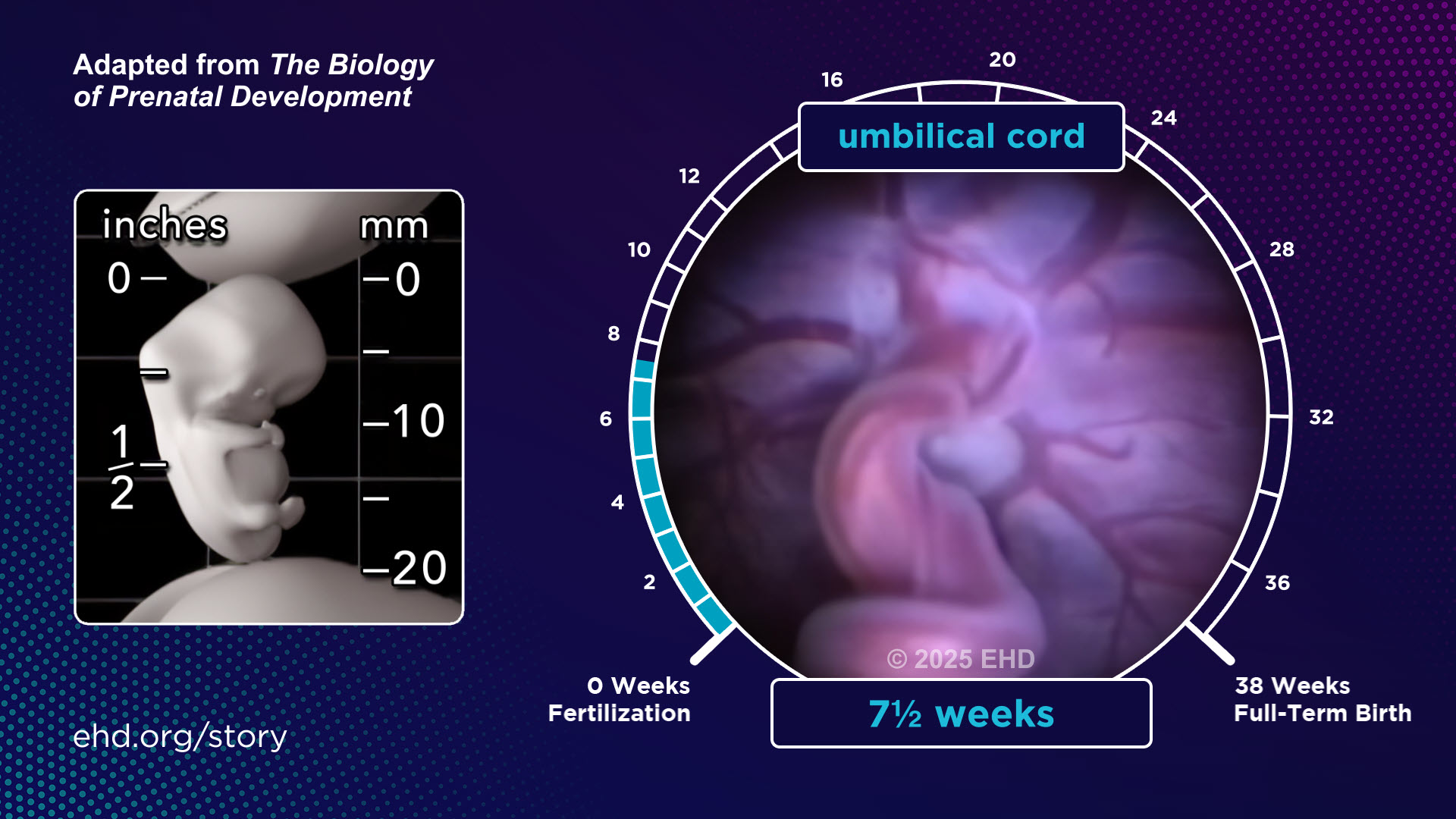
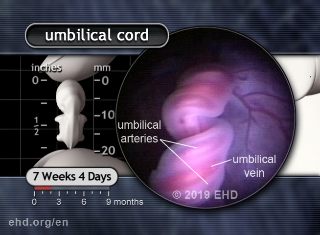
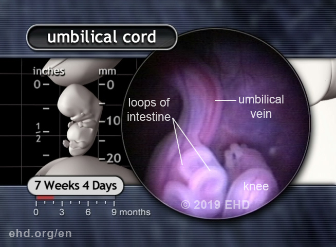
The Amazing Placenta and Umbilical Cord
The placenta serves as the vital interface between mother and child. It produces hormones, helps regulate the baby's body temperature, supplies oxygen, removes carbon dioxide, and more.
Placental origin of umbilical cord
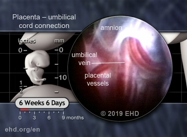
The origin of the umbilical cord
Placenta from the inside
Umbilical cord details
The origin of rhythm
Base of the umbilical cord
Footnotes