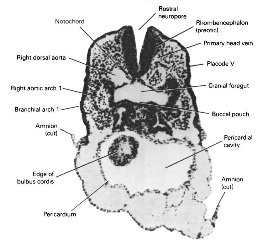
A section through the first branchial arch and the cranial part of the pericardial cavity.
Observe:
1. Because of the head fold (see Figs. 4–1 and 4–2B), the attachment of the amnion moves to the ventral side of the head region.
2. The cranial edge of the primitive bulbus cordis surrounded by the pericardium on the ventral aspect of the head.
3. Within the first branchial arch is the first aortic arch about to join the dorsal aorta.
4. The slight ventral bulge in the floor of the foregut forming the buccal pouch.
5. The first appearance of the primary head vein between the brain and dorsal aorta.
Keywords: cephalic neuropore, cephalic part of foregut, cut edge of amnion, edge of bulbis cordis, notochord, pericardial cavity, pericardial sac, pharyngeal arch 1, placode 5, primary head vein, rhombencephalon (preotic), right aortic arch 1, right dorsal aorta, vestibule of oral cavity
Source: Atlas of Human Embryos.