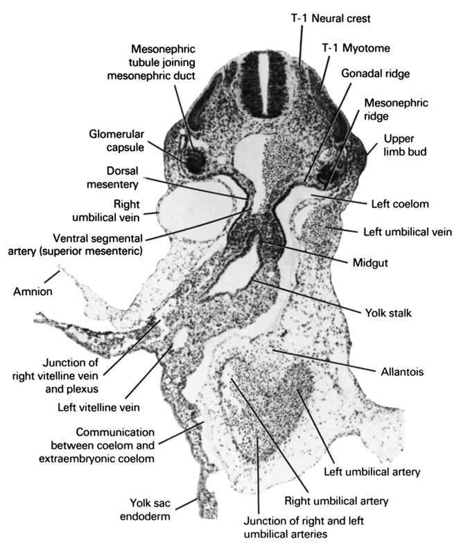
A section through the T-1 neural crest and the middle of the midgut and yolk stalk junction.
Observe:
1. The junction of the right and left umbilical arteries.
2. The cranial edge of the allantois.
3. The attachment of the amnion to the ventral body wall.
4. The vitelline artery that is a continuation of one of the ventral segmental arteries.
5. A mesonephric tubule joining its glomerular capsule with the mesonephric duct.
Keywords: T-1 myotome, T-1 neural crest, allantois, amnion, communication between coelom and extraembryonic coelom, dorsal mesentery, glomerular capsule, gonadal ridge, junction of right and left umbilical arteries, junction of right vitelline vein and plexus, left coelom, left umbilical artery, left umbilical vein, left vitelline (omphalomesenteric) vein, mesonephric ridge, mesonephric tubule joining mesonephric duct, midgut, right umbilical artery, right umbilical vein, upper limb bud, ventral segmental artery (superior mesenteric), yolk sac endoderm, yolk stalk
Source: Atlas of Human Embryos.