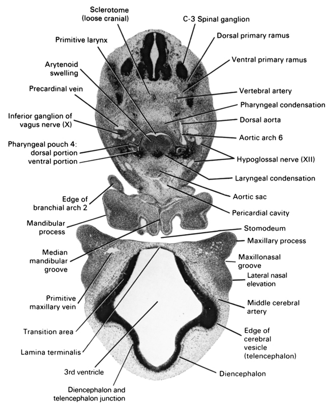
A section through the primitive larynx and fourth pharyngeal pouch.
Observe:
1. The dorsal and ventral primary rami of the C-3 spinal ganglion.
2. The dorsal portion of the fourth pharyngeal pouch that gives rise to the rostral (superior) parathyroid gland and the ventral portion that forms the lesser thymus.
3. The arytenoid swelling on each side of the primitive larynx.
4. The lamina terminalis and the di- and telencephalon junction.
5. The maxillary process and lateral nasal elevation separated by the maxillonasal groove.
Keywords: C-3 spinal ganglion, aortic arch 6, aortic sac, arytenoid swelling, diencephalon, diencoel (third ventricle), dorsal aorta, dorsal portion of pharyngeal pouch 4, dorsal primary ramus, edge of cerebral vesicle (telencephalon), edge of pharyngeal arch 2, hypoglossal nerve (CN XII), inferior ganglion of vagus nerve (CN X), junction of diencephalon and telencephalon, lamina terminalis, laryngeal condensation, lateral nasal elevation, mandibular process, maxillary process, maxillonasal groove, median mandibular groove, middle cerebral artery, pericardial cavity, pharyngeal condensation, precardinal vein, primitive larynx, primitive maxillary vein, sclerotome (loose cranial), stomodeum, transition area, ventral portion of pharyngeal pouch 4, ventral primary ramus, vertebral artery
Source: Atlas of Human Embryos.
