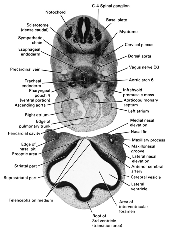
A section through the C-4 spinal ganglion and the cranial edge of the heart.
Observe:
1. The separation of the pharyngeal endoderm into esophageal and tracheal portions.
2. The infrahyoid premuscle mass on either side of the heart in the cervical region.
3. The right and left atria separated by the ascending aorta and pulmonary trunk.
4. The edge of the nasal pit with the nasal fin separating the medial nasal elevation from the maxillary process.
5. The two subdivisions of the telencephalon: cerebral vesicle and telencephalon medium.
Keywords: C-4 spinal ganglion, anterior cerebral artery, aortic arch 6, aorticopulmonary septum, area of interventricular foramen, ascending aorta, basal plate, cerebral vesicle, cervical plexus, dorsal aorta, edge of nasal pit, edge of pulmonary trunk, esophageal endoderm, infrahyoid premuscle mass, lateral nasal elevation, lateral ventricle, left atrium, maxillary process, maxillonasal groove, medial nasal elevation, myotome, nasal fin, notochord, pericardial cavity, pharyngeal pouch 4, pre-optic area, precardinal vein, right atrium, roof of third ventricle (transition area), sclerotome (dense caudal), striatal part, suprastriatal part, sympathetic chain, telencephalon medium, tracheal endoderm, vagus nerve (CN X)
Source: Atlas of Human Embryos.