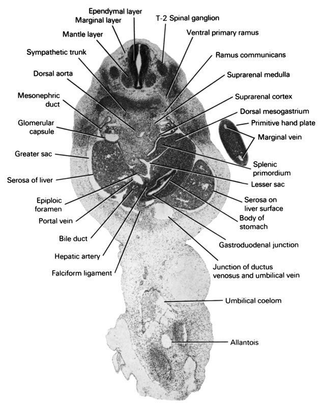
A section through the T-2 spinal ganglion, suprarenal gland and gastroduodenal junction.
Observe:
1. A ramus communicans connecting the sympathetic trunk with a ventral primary ramus.
2. The splenic primordium in the dorsal mesogastrium.
3. The lesser sac communicating through the epiploic foramen with the greater sac.
4. The portal vein, bile duct and hepatic artery ventral to the epiploic foramen.
5. The junction of the ductus venosus and umbilical vein at the liver surface.
Keywords: T-2 spinal ganglion, allantois, bile duct, body of stomach, dorsal aorta, dorsal mesogastrium, ependymal layer, epiploic foramen, falciform ligament, gastroduodenal junction, glomerular capsule, greater sac, hepatic artery, junction of ductus venosus and umbilical vein, lesser sac, mantle layer, marginal layer, marginal vein, mesonephric duct, portal vein, primitive hand plate, ramus communicans, serosa of liver, serosa on liver surface, splenic primordium, suprarenal cortex, suprarenal medulla, sympathetic trunk, umbilical coelom, ventral primary ramus
Source: Atlas of Human Embryos.