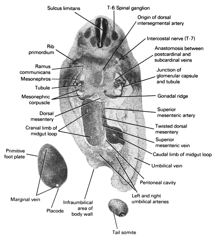
A section through the T-6 spinal ganglion and junction of the umbilical vessels with the ventral body wall.
Observe:
1. The position of the intercostal nerve to the dorsal end of the rib primordium.
2. The anastomosis between the post- and subcardinal veins medial to the mesonephros.
3. The junction of a glomerular capsule and tubule in the mesonephros.
4. The superior mesenteric vessels in the twisted dorsal mesentery of the midgut.
5. The marginal vein and placode of the primitive foot plate.
Keywords: T-6 spinal ganglion, anastomosis between postcardinal and subcardinal veins, caudal limb of midgut loop, cranial limb of midgut loop, dorsal mesentery, gonadal ridge, infraumbilical area of body wall, intercostal nerve (T-7), junction of glomerular capsule and tubule, left and right umbilical arteries, marginal vein, mesonephric corpuscle, mesonephros, origin of dorsal intersegmental artery, peritoneal cavity, placode, primitive foot plate, ramus communicans, rib primordium, sulcus limitans, superior mesenteric artery, superior mesenteric vein, tail somite, tubule, twisted dorsal mesentery, umbilical vein
Source: Atlas of Human Embryos.