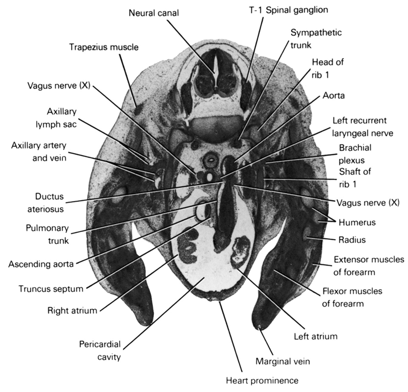
A section through the T-1 spinal ganglion and cranial edge of the atria.
Observe:
1. The ascending aorta to the right of the pulmonary trunk from which it is separated by the truncus septum.
2. The ductus arteriosus connecting the pulmonary trunk and aorta.
3. The left vagus nerve lateral to the ductus arteriosus with its branch, the recurrent laryngeal nerve, medial.
4. The sympathetic trunk medial to the head of the first rib.
5. The relative position of the extensor and flexor muscles in the forearm.
Keywords: T-1 spinal ganglion, aorta, ascending aorta, axillary artery, axillary lymph sac, axillary vein, brachial plexus, ductus arteriosus, extensor muscles of forearm, flexor muscles of forearm, head of rib 1, heart prominence, humerus, left atrium, left recurrent laryngeal nerve, marginal vein, neural canal, pericardial cavity, pulmonary trunk, radius, right atrium, shaft of rib 1, sympathetic trunk, trapezius muscle, truncus septum, vagus nerve (CN X)
Source: Atlas of Human Embryos.