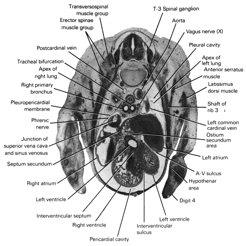
A section through the T-3 spinal ganglion and the tracheal bifurcation.
Observe:
1. The cranial part of the left ventricle dorsal to the right ventricle from which it is separated by the membranous part of the interventricular septum.
2. The septum secundum between the right and left atria and the ostium secundum through which the two atria communicate.
3. The entrance of the superior vena cava into the pericardial cavity and its junction with the sinus venosus.
4. The phrenic nerve coursing through the thin pleuropericardial membrane that separates the pericardial and pleural cavities.
5. The primordium of the fourth digit of the hand.
Keywords: T-3 spinal ganglion, anterior serratus muscle, aorta, apex of left lung, apex of right lung, atrioventricular sulcus, digit 4, erector spinae muscle group, hypothenar area, interventricular septum, interventricular sulcus, junction of superior vena cava and sinus venosus, latissimus dorsi muscle, left atrium, left common cardinal vein, left ventricle, ostium secundum area, pericardial cavity, phrenic nerve, pleural cavity, pleuropericardial membrane, postcardinal vein, right atrium, right primary bronchus, right ventricle, septum secundum, shaft of rib 3, tracheal bifurcation, transversospinal muscle group, vagus nerve (CN X)
Source: Atlas of Human Embryos.