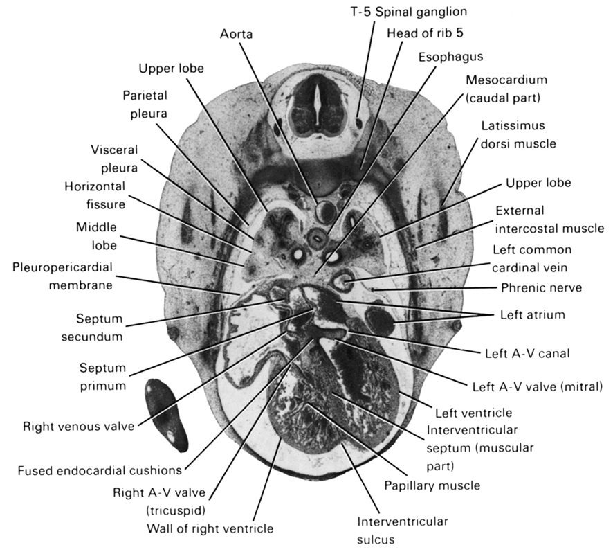
A section through the T-5 spinal ganglion and the right and left atrioventricular valves.
Observe:
1. A papillary muscle attaching a leaf of the right atrioventricular valve to the wall of the right ventricle.
2. The septum primum caudal to the ostium secundum (see Section 31) and left of the septum secundum.
3. The right venous valve joining the fused endocardial cushions.
4. The left atrioventricular canal connecting the left atrium with the left ventricle.
5. The esophagus passing ventral to the aorta.
Keywords: T-5 spinal ganglion, aorta, esophagus, external intercostal muscle(s), fused endocardial cushions, head of rib 5, horizontal fissure, interventricular septum (muscular part), interventricular sulcus, latissimus dorsi muscle, left atrioventricular (mitral) valve, left atrioventricular canal, left atrium, left common cardinal vein, left ventricle, mesocardium (caudal part), middle lobe, papillary muscle, parietal pleura, phrenic nerve, pleuropericardial membrane, right atrioventricular (tricuspid) valve, right venous valve, septum primum, septum secundum, upper lobe, visceral pleura, wall of right ventricle
Source: Atlas of Human Embryos.