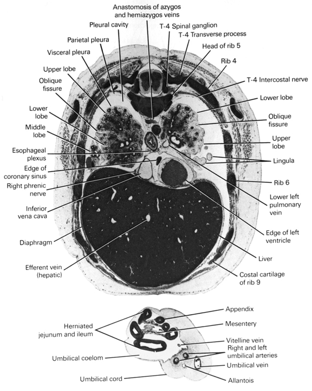
A section through the umbilical cord, caudal edge of the heart and the T-4 spinal ganglion.
Observe:
1. The blood vessels, allantois and herniated gut in the umbilical cord.
2. The right phrenic nerve to the right of the inferior vena cava at the diaphragm.
3. The esophageal plexus formed by the vagus nerves.
4. The oblique fissure in the left lung separating the upper lobe with its lingula from the lower lobe.
5. The parietal and visceral subdivisions of the pleura lining the pleural cavity.
Keywords: T-4 intercostal nerve, T-4 spinal ganglion, T-4 transverse process, allantois, anastomosis between azygos and hemi-azygos veins, appendix, costal cartilage of rib 9, diaphragm, edge of coronary sinus, edge of left ventricle, efferent vein (hepatic), esophageal nerve plexus, head of rib 5, herniated jejunum and ileum, inferior vena cava, lingula, liver, lower left pulmonary vein, lower lobe of left lung, lower lobe of right lung, mesentery, middle lobe of right lung, oblique fissure, parietal pleura, pleural cavity, rib 4, rib 6, right and left umbilical arteries, right phrenic nerve, umbilical coelom, umbilical cord, umbilical vein, upper lobe of left lung, upper lobe of right lung, visceral pleura, vitelline (omphalomesenteric) vein
Source: Atlas of Human Embryos.
