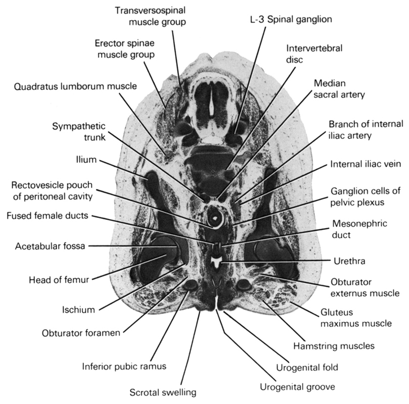
A section through the urogenital groove and fold and L-3 spinal ganglion.
Observe:
1. The three cartilagenous precursors of the pelvic bone; ilium, ischium and pubis.
2. The near junction of the mesonephric ducts with the urethra.
3. The fused female ducts in the midline.
4. The rectovesicle pouch of the peritoneal cavity.
5. The ganglion cells of the pelvic plexus lateral to the pelvic viscera.
Keywords: L-3 spinal ganglion, acetabular fossa, branch of internal iliac artery, erector spinae muscle group, fused female ducts, ganglion cells of pelvic plexus, gluteus maximus muscle, hamstring muscles, head of femur, ilium, inferior pubic ramus, internal iliac vein, intervertebral disc, ischium, median sacral artery, mesonephric duct, obturator externus muscle, obturator foramen, quadratus lumborum muscle, rectovesicle pouch of peritoneal cavity, scrotal swelling, sympathetic trunk, transversospinal muscle group, urethra, urogenital fold, urogenital groove
Source: Atlas of Human Embryos.