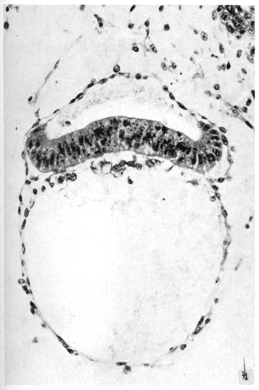
The embryo and secondary umbilical vesicle from the same section are shown more highly magnified in this figure. At its turned-up margins, the ectodermal plate changes abruptly to the thin squamous cells of the amnion. The foamy nature of the gut endoderm, in contrast with the flattened endodermal cells lining the secondary umbilical vesicle, is apparent. The external coat of the secondary umbilical vesicle is a thin layer closely applied to the lining endoderm. The position of the section in the embryo is indicated in figure 13. Section 135.
Fig. 4. Heuser et al., 1945.
Click on the picture to view the full-sized image.
Keywords: amnion, ectodermal plate, gut endoderm, secondary umbilical vesicle
Source: The Virtual Human Embryo.