Skip Navigation

Carnegie Stage Section
 <   >  search results: optic stalk lumen (CN II) Search   14-320 14-329 14-338 16-345 16-355 17-337 17-343 17-371
C-3 spinal ganglion, aortic arch 3, arytenoid swelling, carotid duct, diencephalon, dorsal sulcus, edge of lens vesicle, epithalamus, hypaxial part of myotome, hypoglossal nerve (CN XII), intraretinal space (optic vesicle cavity), junction of intraretinal space (optic vesicle cavity) and optic stalk lumen (CN II), neural layer of retina, optic chiasma (chiasmatic plate), optic stalk lumen (CN II), pigmented layer of retina, pineal bud, precardinal vein, premuscle mass of tongue, retinal fissure, terminal branches of mandibular nerve (CN V₃), thyroglossal duct, ventral part of pharyngeal pouch 3 becomes thymus gland, vertebral artery
0 μm
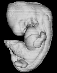
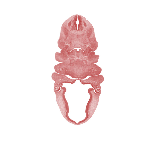
Carnegie Embryo #6517   |   Location: 15-04-01

Keywords: C-3 spinal ganglion, aortic arch 3, arytenoid swelling, carotid duct, diencephalon, dorsal sulcus, edge of lens vesicle, epithalamus, hypaxial part of myotome, hypoglossal nerve (CN XII), intraretinal space (optic vesicle cavity), junction of intraretinal space (optic vesicle cavity) and optic stalk lumen (CN II), neural layer of retina, optic chiasma (chiasmatic plate), optic stalk lumen (CN II), pigmented layer of retina, pineal bud, precardinal vein, premuscle mass of tongue, retinal fissure, terminal branches of mandibular nerve (CN V₃), thyroglossal duct, ventral part of pharyngeal pouch 3 becomes thymus gland, vertebral artery

Source: The Virtual Human Embryo.