د زېږېدنې څخه مخکې د ماشوم د ودې بيالوژي
The Biology of Prenatal Development
DVD Documentation
English / پښتو [Pashtu (Pushto)]
Show Script Cover & Table of Contents
English / پښتو [Pashtu (Pushto)]
Chapter 1 Introduction
The dynamic process by which the single-cell human zygote(zī΄gōt)[1] becomes a 100 trillion (1014) cell adult[2] is perhaps the most remarkable phenomenon in all of nature.
Researchers now know that many of the routine functions performed by the adult body become established during pregnancy – often long before birth.[3]
The developmental period before birth is increasingly understood as a time of preparation during which the developing human acquires the many structures, and practices the many skills, needed for survival after birth.
Chapter 1 Introduction
هغه خوځنده پروسه، چې په هغې د انسان يو حجروي زايګوټ په يو ۱۰۰ تريليون- حجروي بالغ انسان باندې بدلېږي چې ښايي په طبيعت کې تر ټولو غوره او د پام وړ پېښه وي.
څېړونکي اوس پوهېږي چې ډېرې عادي دندې چې د بالغ انسان بدن په واسطه سرته رسېږي د اُميندوارۍ (حامله ګۍ) په موده کې يې بنسټ ايښودل کېږي، چې زياتره تر زېږېدنې ډېر وړاندې تر سره کيږي .
له زېږېدنې څخه مخکې د ودې موده، دچمتو کولو دوخت په توګه پېژ ندل کيږ ي. هغه مهال چې د ودې په حال کې انسان ګڼ شمېر جوړښتونه، تمرينونه اوګڼ شمېر مهارتونه چې ورته اړتيا لري له زېږېدنې ورسته د پايښت لپاره ترلاسه کوي.
Chapter 2 Terminology
Pregnancy in humans normally lasts approximately 38 weeks[4] as measured from the time of fertilization,[5] or conception,[6] until birth.
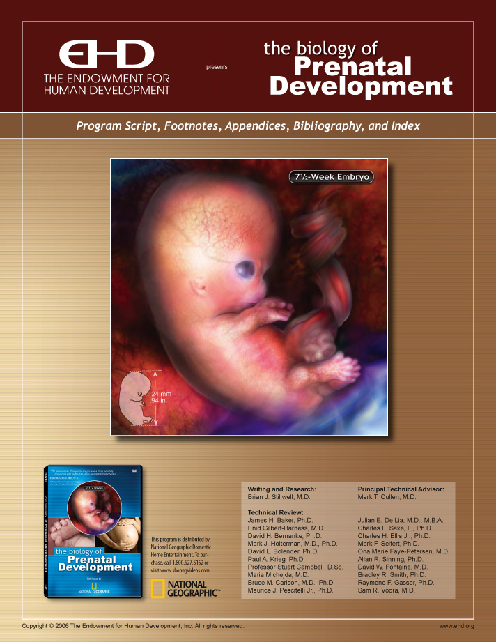
During the first 8 weeks following fertilization, the developing human is called an embryo,[7] which means "growing within."[8] This time, called the embryonic period,[9] is characterized by the formation of most major body systems.[10]
From the completion of 8 weeks until the end of pregnancy, "the developing human is called a fetus," which means "unborn offspring." During this time, called the fetal period, the body grows larger and its systems begin to function.[11]
All embryonic and fetal ages in this program refer to the time since fertilization.[12]
Chapter 2 Terminology
په انسانانو کې د اميدوارۍ (موده) په عادي ډول تقريباً ۳۷ اوونۍ دوام کوي. چې د القاح له وخت څخه اندازه شوى يا له اميدوارۍ نه، تر زېږېدنې پورې.
د القاح پسې د لومړيو ۸ اوونيو په موده کې، د ودې په حال کې انسان ته امبريو (جنين) وايي، چې معنا يې ده "دننه وده کول" دغه وخت چې د جنين د دورې په نامه يادېږي، د تشکل په نامه ځانګړې شوې چې، د بدن د ډېرو عمده سيستمونود تشکل دوره ده.
د اتو اوونيو له پوره کېدو څخه د اميدوارۍ تر پايه پورې، د ودې په حال انسان ته کامل شوی جنين (فيټس) وايي، چې معنا يې "نازېږېدلی ماشوم" دى. په دغه موده کې چې د جنين (فيټال) دوران په نامه يادېږي، بدن د لويېدو په لور وده کوي او غړي يې په فعاليت پېل کوي.
په دې پروګرام کې ټول جنين او د کامل شوي جنين عمرونه هغه وخت ته رجوع کوي چې له القاح څخه شروع کېږي.
Click any superscript in the text to view footnote. Click any footnote number to view source text. Click on any author name to view the full reference in the Bibliography. Then click your browser’s back button to return to source footnote.
[1]
Gasser, 1975, 1.
[2]
Guyton and Hall, 2000, 2;
Lodish et al., 2000, 12.
[3]
Vindla and James, 1995, 598.
[4]
Cunningham et al., 2001, 226;
O’Rahilly and Müller, 2001, 92.
[5]
O’Rahilly and Müller, 1987, 9.
[6]
Spraycar, 1995, 377 & 637.
[7]
O’Rahilly and Müller, 2001, 87.
[8]
Quote from Ayto, 1990, 199.
[9]
Human development during the 8-week embryonic period has been divided into a series of 23 stages called Carnegie Stages. These stages are well described in O’Rahilly and Müller, 1987. Because human growth is unique and dependent on multiple factors, different embryos may reach a certain developmental milestone or a certain size at slightly different ages. This internationally-accepted staging system provides a way to describe development independent of age and size. Each of the 23 Carnegie Stages has specific structural features. As we describe various milestones of development, the Carnegie Stage at which they occur will be noted by a designation such as: [Carnegie Stage 2]. See Appendix B for additional information relating embryonic staging and age assignments.
[10]
Moore and Persaud, 2003, 3.
[11]
Quotes from Moore and Persaud, 2003, 3: “After the embryonic period (eight weeks), the developing human is called a fetus.“ Also see O’Rahilly and Müller, 2001, 87.
[12]
This convention, termed “postfertilization age“ by O’Rahilly, has been long preferred by embryologists. [see Mall, 1918, 400;
O’Rahilly and Müller, 1999b, 39;
O’Rahilly and Müller, 2001, 88 & 91.] Obstetricians and radiologists typically assign age based on the time elapsed since the first day of the last menstrual period prior to fertilization. This is correctly termed “postmenstrual age“ and begins 2 weeks before fertilization occurs. To summarize: postmenstrual age = postfertilization age + 2 weeks. Therefore, postmenstrual age equals approximately 2 weeks at the time of fertilization. The commonly used term “gestational age“ has been used with both age conventions and is best either avoided or carefully defined with each use.
Page 3
The Embryonic Period (The First 8 Weeks)
Embryonic Development: The First 4 Weeks
Chapter 3 Fertilization
Biologically speaking, "human development begins at fertilization,"[13] when a woman and a man each combine 23 of their own chromosomes through the union of their reproductive cells.
A woman's reproductive cell is commonly called an "egg" but the correct term is oocyte (ō´ō-sīt).[14]
Likewise, a man's reproductive cell is widely known as a "sperm," but the preferred term is spermatozoon (sper´mă-tō-zō´on).[15]
Following the release of an oocyte from a woman's ovary in a process called ovulation (ov´yū-lā´shŭn),[16] the oocyte and spermatozoon join within one of the uterine tubes,[17] which are often referred to as Fallopian tubes.
The uterine tubes link a woman's ovaries to her uterus or womb.
The resulting single-celled embryo is called a zygote,[18] meaning "yoked or joined together."[19]
The Embryonic Period (The First 8 Weeks)
Embryonic Development: The First 4 Weeks
Chapter 3 Fertilization
که په بيالوژيکي توګه خبرې وکړو، "د انسان وده د القاح له وخته پېل کېږي،" کله چې يوه ښځه او يو سړی هر يو خپل ۲۳ کروموزومونه يوځای کړي د خپلو تکثري ژوانکو( حجرو) د يو ځاى کولو له لارې.
د ښځې تکثري ژوآنکه( حجره) په عامه توګه د هګۍ په نامه يادېږي، خو سمه اصطلاح يې اووسايټ دى..
همدارنګه، د يو سړي تکثري ژوانکې (حجرې) په پراخه اندازه د سپرم(مني) په نامه پېژندل شوي خو سمه اصطلاح يې سپرماتوزون ده.
د ښځې له تخمدان څخه د نا القاح شوې هګۍ د راوتو څخه وروسته په هغه پروسه کې چې دهګې توليدولو(اوويوليشن) په نامه يادېږي، د ښځې هګۍ (اووسايټ) او د نارينه سپرم (مني) (سپرماتوزون) يوځای کېږي (دا دواړه) د زهدان (رحم) نلو څخه په يوه کې، چې زياتره ورته (فالوپين) ټيوبونه هم وايي، (يوځاى کېږي).
زهداني ټيوب (د رحم نل) د ښځې تخمدانونه د هغې له زهدان (رحم) سره يوځای کوي
په نتيجه کې چې کوم يو حجروي جنين مينځ ته راځې د زايګوټ په نامه يادېږي، چې معنا يې جوړه کيدل يا يوځای کېدل دي.
Chapter 4 DNA, Cell Division, and Early Pregnancy Factor (EPF)
DNA
The zygote's 46 chromosomes[20] represent the unique first edition of a new individual's complete genetic blueprint. This master plan resides in tightly coiled molecules called DNA. They contain the instructions for the development of the entire body.
DNA molecules resemble a twisted ladder known as a double helix.[21] The rungs of the ladder are made up of paired molecules, or bases, called guanine, cytosine, adenine, and thymine.
Guanine pairs only with cytosine, and adenine with thymine.[22] Each human cell contains approximately 3 billion (3×10⁹) base pairs.[23]
The DNA of a single cell contains so much information that if it were represented in printed words, simply listing the first letter of each base would require over 1.5 million (1.5×10⁶) pages of text![24]
If laid end-to-end, the DNA in a single human cell measures 3⅓ feet or 1 meter.[25]
If we could uncoil all of the DNA within an adult's 100 trillion (1014) cells, it would extend over 63 billion (6.3×10¹⁰) miles. This distance reaches from the earth to the sun and back 340 times.[26]
Cell Division
Approximately 24 to 30 hours after fertilization, the zygote completes its first cell division.[27] Through the process of mitosis, one cell splits into two, two into four, and so on.[28]
Early Pregnancy Factor (EPF)
As early as 24 to 48 hours after fertilization begins, pregnancy can be confirmed by detecting a hormone called "early pregnancy factor" in the mother's blood.[29]
Chapter 4 DNA, Cell Division, and Early Pregnancy Factor (EPF)
DNA
د زايګوټ ۴۶ کروموزمونه لومړنۍ بې سارې لړۍ تمثيلوي دا د نوي شخص بشپړه جنيتکي طرحه ده. دغه عمومي پلان موقعيت لري (يعنې )په کلکو تاوشويو ماليکولونو کې چې د ډي اين ای په نامه يادېږي. دوى د ودې لپاره لارښوونې لري د ټول بدن.
د ډي اين ای ماليکولونه يوې تاوراتاو شوې زينې ته ورته دي چې د غبرګ فنر (ډبل هيليکس) په نامه يادېږي . د زينې د پاټکيو ميلې له غبرګو ماليکولونو څخه جوړې شوې دي يا القلي څخه چې عبارت دي له ګوانين، سايتوزين، اډينين او تايمين.
ګوانين يواځې له سايتوزين سره جوړه کېږي ، او اډينين له تايمين سره. د انسان هره ژوانکه (حجره )تقريباً ٣ بليونه لري. (يعنې )له دې بنسټيزو جوړو څخه.
د يوې واحدې ژوانکې (حجرې) ډي اين ای د ډېرو زياتو معلوماتو لرونکى دى که دغه معلومات په چاپ شويو کلمو کې ښودل شوي وای، او په ساده ډول د هر بنسټ ،لومړی توری ليست شوى واى. نو متن ته به له ١.٥ مليون پاڼو څخه زياتو ته اړتيا واى!
که څوکه په څوکه پرله پسې کېښودل شي، د انساني واحدې ژوانکې د ډي اين اې اندازه به ۳/۳۱ فټه يا يو مترواى.
که مونږ د ډي اين اې ټولې حلقې خلاصې کړای شو په يو بالغ(انسان) کې۱۰۰ تريلىونه ژوانکې )، به له ۶۳ بيليونه ميلو څخه زياتې اوږدې شي. چې دغه واټن له ځمکې تر لمر او له لمر څخه بېرته ځمکې ته ۳۴۰ ځلې ته رسېږي.
Cell Division
له القاح څخه،تقريباً ۲۴ نه تر ۳۰ ساعته وروسته زايګوټ خپل لومړنى حجروي وېش تکميلوي د مايتوسس د پروسې له لارې، يوه ژوانکه پر دوه و، دوه پر څلورو او په همدې ترتيب وېشل کېږي.
Early Pregnancy Factor (EPF)
د القاح نه لږ تر لږه له ٢٤ ساعتو تر ٤٨ ساعتو وروسته د يو هارمون په ترشح کېدو حامله داري تصديق کيداى شي. چې د مور په وينه کې د وختي حامله دارۍ د عامل په نامه يادېږي.
[13]
Quote from Moore and Persaud, 2003, 16;
From O’Rahilly and Müller, 1987, 9: “Fertilization is the procession of events that begins when a spermatozoon makes contact with an oocyte or its investments and ends with the intermingling of maternal and paternal chromosomes at metaphase of the first mitotic division of the zygote.“ See Carlson, 2004, 3;
O’Rahilly and Müller, 2001, 8. [Carnegie Stage 1]
[14]
O’Rahilly and Müller, 2001, 25: “The term ‘egg’ should be discarded from human embryology.“ From O’Rahilly and Müller, 1987, 9: “The term ‘egg’ is best reserved for a nutritive object frequently seen on the breakfast table.“
[15]
O’Rahilly and Müller, 2001, 23-24.
[16]
O’Rahilly and Müller, 2001, 30.
[17]
Dorland and Bartelmez, 1922, 372;
Gasser, 1975, 1;
Mall, 1918, 421;
O’Rahilly and Müller, 2001, 31.
[18]
Gasser, 1975, 1;
O’Rahilly and Müller, 2001, 33.
[19]
Quote from Saunders, 1970, 1;
Spraycar, 1995, 1976.
[20]
Guyton and Hall, 2000, 34.
[21]
Guyton and Hall, 2000, 24;
Watson and Crick, 1953, 737.
[22]
Guyton and Hall, 2000, 24;
Lodish et al., 2000, 103;
Watson and Crick, 1953, 737.
[23]
Lodish et al., 2000, 456.
[24]
See Appendix A.
[25]
See Appendix A;
Alberts et al., 1998, 189.
[26]
See Appendix A.
[27]
Hertig, 1968, 26;
Hertig and Rock, 1973, 130;
(cited by O’Rahilly and Müller, 1987, 12);
Shettles, 1958, 400.
[28]
Guyton and Hall, 2000, 34.
[29]
Moore and Persaud, 2003, 33 & 60;
Morton et al., 1992, 72;
Nahhas and Barnea, 1990, 105.
Page 4
Chapter 5 Early Stages (Morula and Blastocyst) and Stem Cells
By 3 to 4 days after fertilization, the dividing cells of the embryo assume a spherical shape and the embryo is called a morula (mōr´ū-lă).[30]
By 4 to 5 days, a cavity forms within this ball of cells and the embryo is then called a blastocyst.[31]
The cells inside the blastocyst are called the inner cell mass and give rise to the head, body, and other structures vital to the developing human.[32]
Cells within the inner cell mass are called embryonic stem cells because they have the ability to form each of the more than 200 cell types contained in the human body.[33]
Chapter 5 Early Stages (Morula and Blastocyst) and Stem Cells
د القاح څخه ۳ - ۴ ورځې وروسته د جنين وېشل کېدونکې ژوانکې بيضوي بڼه غوره کوي او جنين د (مارولا) په نامه يادېږي.
۴ - ۵ ورځو کې د ژوانکو د پنډوسکې په منځ کې يوچوغاړى جوړېږي او جنين د (بلاستوسيسټ) په نامه يادېږي.
په بلاستوسيسټ کې دننه حجرې د داخلي حجرو د کتلې په نامه يادېږي او وده ورکوي سر، تنې او د بدن نورو غړو ته چې د ودې په حال انسان ته ډېر ضروري دي .
ژوانکې په دننني حجروي کتله کې د جنيني تنې د ژوانکو په نامه يادېږي ځکه دوی ددې وړتيا لري چې هر يو له ۲۰۰ ډولو ژوانکو څخه زيات تشکيل کړي چې انساني بدن يې لري.
Chapter 6 1 to 1½ Weeks: Implantation and Human Chorionic Gonadotropin (hCG)
After traveling down the uterine tube, the early embryo embeds itself into the inner wall of the mother's uterus. This process, called implantation, begins 6 days and ends 10 to 12 days after fertilization.[34]
Cells from the growing embryo begin to produce a hormone called human chorionic gonadotropin (human kō-rē-on'ik gō'nad-ō-trō'pin), or hCG, the substance detected by most pregnancy tests.[35]
HCG directs maternal hormones to interrupt the normal menstrual cycle, allowing pregnancy to continue.[36]
Chapter 6 1 to 1½ Weeks: Implantation and Human Chorionic Gonadotropin (hCG)
د رحم په نل کې له تېرېدو وروسته، لومړنى زايګوټ خپل ځان محاطوي. د مور د رحم په داخلي دېوال کې . دغه پروسه، چې د غرس کولو (امپلانټېشن) په نامه يادېږي، په ۶ ورځو کې پېل کېږي او له القاح څخه وروسته له ۱۰ نه تر ۱۲ ورځو پورې پای ته رسېږي.
د ودې په حال کې جنيني ژوانکې د يو هارمون په جوړولو پېل کوي چې د انساني ـ کوريونيک ګونادوټروپين ـ يا ايچ. سي. جي.په نامه يادېږي، چې د حاملګۍ د ډېرو ټسټونو په واسطه تشخېصېږي.
ايچ. سي. جي. مورني هارمونونه لارښوونه کوي چی د حيض په نورمال دوران کې مداخله وکړي، او اجازه ورکوي چې حاملګي دوام وکړي
Chapter 7 The Placenta and Umbilical Cord
Following implantation, cells on the periphery of the blastocyst give rise to part of a structure called the placenta (plă-sen'tă), which serves as an interface between the maternal and embryonic circulatory systems.
The placenta delivers maternal oxygen, nutrients, hormones, and medications to the developing human; removes all waste products; and prevents maternal blood from mixing with the blood of the embryo and fetus.[37]
The placenta also produces hormones and maintains embryonic and fetal body temperature slightly above that of the mother's.[38]
The placenta communicates with the developing human through the vessels of the umbilical (ŭm-bil'i-kăl) cord.[39]
The life support capabilities of the placenta rival those of intensive care units found in modern hospitals.
Chapter 7 The Placenta and Umbilical Cord
د غرس کولو په تعقيب هغه حجرې چې د بلاسټوسايټ په بهرنۍ سطح کې دي د يو جوړښت يوې برخې ته وده ورکوي چې د پلاسنټا په نامه يادېږي. هغه چې د مورني ګډ ټکي په څېر خدمت کوي. اود جنيني وينې د دوران سيستمونو کې.
پلاسنټا، مورنى اکسيجن، خوراک، هارمونونه او درملونه دودې په حال انسان ته وروېشي ; فضله مواد وباسي ; او د مورنۍ وينې له ګډېدلو څخه مخنيوی کوي (يعنې) له وينې د جنين او د نازيږيدلي ماشوم (فيټس) سره.
پلاسنټا هم هارمونونه توليدوي د جنيني او د نازېږېدلي ماشوم د بدن تودوخه ثابته ساتي د مور له حرارت څخه لوړ
پلاسنټا د ودې په حال انسان سره ارتباط نيسي د نامه مزى د رګونو له لارې.
د ژوند د سا تنې استعداد چې پلاسنټا يې لري د عصري روغتونو د سختو ساتندويه دستګاو(آی. سي يو) سره ورته والی لري.
[30]
Gasser, 1975, 1;
O’Rahilly and Müller, 2001, 37;
Spraycar, 1995, 1130: “Morula“ is derived from the Latin word morus meaning “mulberry.“ [Carnegie Stage 2]
[31]
O’Rahilly and Müller, 2001, 39. [Carnegie Stage 3]
[32]
Gasser, 1975, 1;
O’Rahilly and Müller, 2001, 39;
Sadler, 2005, 6.
[33]
Alberts et al., 1998, 32. For a discussion and definition of embryonic stem cells see the website of the National Institutes of Health: http://stemcells.nih.gov/infoCenter/stemCellBasics.asp#3
[34]
O’Rahilly and Müller, 2001, 40;
Implantation begins with attachment of the blastocyst at about 6 days after fertilization. [Attachment of the blastocyst to the inner wall of the uterus is a transient event and is the hallmark of Carnegie Stage 4.] See also Adams, 1960, 13-14;
Cunningham et al., 2001, 20;
Hamilton, 1949, 285-286;
Hertig, 1968, 41;
Hertig and Rock, 1944, 182;
Hertig and Rock, 1945, 81 & 83;
Hertig and Rock, 1949, 183;
Hertig et al., 1956, 444. [Carnegie Stage 5]
[35]
Chartier et al., 1979, 134;
Cunningham et al., 2001, 27;
O’Rahilly and Müller, 2001, 43.
[36]
Cunningham et al., 2001, 20 & 26-27;
O’Rahilly and Müller, 2001, 31.
[37]
Hertig, 1968, 16;
Cunningham et al., 2001, 86 & 136;
For a detailed description of the placenta see Hamilton and Boyd, 1960. For a detailed description of the placenta vasculature see Harris and Ramsey, 1966. This separation of maternal and fetal blood is almost but not quite perfect as a
small number of fetal cells may be found in the maternal circulation and vice-versa. See Cunningham et al., 2001, 96 & 136.
[38]
Liley, 1972, 101;
O’Rahilly and Müller, 2001, 78-79.
[39]
For a detailed description of umbilical cord formation see Florian, 1930.
Page 5
Chapter 8 Nutrition and Protection
By 1 week, cells of the inner cell mass form two layers called the hypoblast and epiblast.[40]
The hypoblast gives rise to the yolk sac,[41] which is one of the structures through which the mother supplies nutrients to the early embryo.[42]
Cells from the epiblast form a membrane called the amnion (am-nē-on),[43] within which the embryo and later the fetus develop until birth.
Chapter 8 Nutrition and Protection
په يوه اوونۍ کې د داخلي حجروي کتلې ژوانکې دوه طبقې جوړوي چې د هايپوبلاسټ او ايپي بلاسټ په نامه يادېږي.
هايپوبلاسټ وده ورکوي د هګۍ د ژېړو (کڅوړې) ته، چې يو له هغو جوړښتونو څخه چې له هغې لارې مور خوراک تهيه کوي. لومړني جنين ته.
(يعنې) د ايپي بلاسټ له حجرو څخه تشکيلېږي يوه غشا چې د امنيون په نامه يادېږي، په جنين کې او وروسته بيا نازېږېدلی ماشوم تر زېږېدلو پورې وده کوي.
Chapter 9 2 to 4 Weeks: Germ Layers and Organ Formation
By approximately 2½ weeks, the epiblast has formed 3 specialized tissues, or germ layers, called ectoderm, endoderm, and mesoderm.[44]
Ectoderm gives rise to numerous structures including the brain, spinal cord, nerves, skin, nails, and hair.
Endoderm produces the lining of the respiratory system and digestive tract and generates portions of major organs such as the liver and pancreas.
Mesoderm forms the heart, kidneys, bones, cartilage, muscles, blood cells, and other structures.[45]
By 3 weeks the brain is dividing into 3 primary sections called the forebrain, midbrain, and hindbrain.[46]
Development of the respiratory and digestive systems is also underway.[47]
As the first blood cells appear in the yolk sac,[48] blood vessels form throughout the embryo, and the tubular heart emerges.[49]
Almost immediately, the rapidly growing heart folds in upon itself as separate chambers begin to develop.[50]
The heart begins beating 3 weeks and 1 day following fertilization.[51]
The circulatory system is the first body system, or group of related organs, to achieve a functional state.[52]
Chapter 9 2 to 4 Weeks: Germ Layers and Organ Formation
تقريباً په ۱/۲ ۲ اوونيو کې، ايپي بلاسټ تشکل موندلى وي. ۳ ځانګړي نسجونه يا د نطفې (جرم) طبقې، چې د ايکټوډرم اينډوډرم ، او ميسوډرم په نامه ياديږي
ايکټوډرم وده ورکوي يو شمېر جو ړښتونو ته چې په کې دماغ، شوکي نخاع ، اعصاب ، پوستکی ، نوکان ، او وېښتان شامل دي.
اينډوډرم د تنفسي سيستم استر او هضمي قنات، او د عمده غړو برخي لکه ځګر (ينه) او تريخى منځ ته راوړي.
له ميزوډرم څخه، زړه، پښتورګي ، هډوکي ، کرپندوکي ، عضلې ، د وينې حجرې، او نور جوړښتونه منځته راځي.
۳ اوونۍ وروسته دماغ په درې لومړنيو برخو وېشل کېږي چې د مخکيني دماغ ، منځني دماغ او شاتني دماغ په نامه يادېږي.
د تنفسي او هضمي جهازونو وده هم پېل شوې ده.
د هګى د زېړو په کڅوړه کې څرګندې شي، د وينې رګونه د جنين په ټولو برخو کې جوړېږي، او نلوزمه زړه راڅرګندېږي.
تقريباً سمدلاسه، په بيړې سره وده کونکی زړه په خپل ځان کې تاو راتاو شي. (يعنې) د بېلابېلو جوفونو په توګه په ودې پېل کوي.
زړه په درځېدو پېل کوي ۳ اوونۍ او يوه ورځ د القاح پسې.
د وينې د دوران سيستم د بدن لومړنی سيستم دی، يا يوه ډله د اړوندو غړو ده، چې فعاله حالت ترلاسه کوي.
Chapter 10 3 to 4 Weeks: The Folding of the Embryo
Between 3 and 4 weeks, the body plan emerges as the brain, spinal cord, and heart of the embryo are easily identified alongside the yolk sac.
Rapid growth causes folding of the relatively flat embryo.[53] This process incorporates part of the yolk sac into the lining of the digestive system and forms the chest and abdominal cavities of the developing human.[54]
Chapter 10 3 to 4 Weeks: The Folding of the Embryo
د ۳ او ۴ اوونيو په منځ کي، د بدن پلان راڅرګندېږي داسې چې دماغ، شوکي نخاع، او د جنين زړه په اسانۍ پېژندل کيږي د هګۍ د زېړو د کڅوړې ترڅنګ.
په بيړه وده د تاوېدو سبب ګرځي (يعنې) نسبتاً پلن جنين. دغه پروسه يوځاې کوي (يعنې) د هګۍ د ژېړو د کڅوړې برخې دننه په استر کې (البته) د هضمې سيستم او سينه تشکل مومي. او بطني خاليګاوې د ودې په حال انسان کې.
[40]
O’Rahilly and Müller, 2001, 39.
[41]
Moore and Persaud, 2003, 50;
O’Rahilly and Müller, 2001, 82. [Carnegie Stages 5 & 6];
In humans, the term “yolk sac“ has fallen out of favor among some embryologists (including O’Rahilly and Müller) because it is not a nutrient reservoir and does not contain yolk. The technically preferred term is umbilical vesicle. This structure plays a vital role in the transfer of nutrients from mother to embryo before placental circulation becomes fully functional.
[42]
Campbell et al., 1993, 756;
Kurjak et al., 1994, 437;
O’Rahilly and Müller, 2001, 82.
[43]
O’Rahilly and Müller, 1987, 29;
O’Rahilly and Müller, 2001, 43. [Carnegie Stages 4-5]
[44]
O’Rahilly and Müller, 2001, 14 & 135. [Carnegie Stage 7];
It should be noted there are many examples of organs derived from multiple germ layers. For instance, the liver is largely derived from endoderm but contains blood vessels and blood cells derived from mesoderm and nerves of ectodermal origin.
[45] Moore
and Persaud, 2003, 80 & 83; Sadler, 2005, 9.
[46]
Bartelmez, 1923, 236;
Müller and O’Rahilly, 1983, 419-420 & 429;
O’Rahilly and Gardner, 1979, 123 & 129;
O’Rahilly and Müller, 1984, 422;
O’Rahilly and Müller, 1987, 90;
O’Rahilly and Müller, 1999a, 47 & 52. [Carnegie Stage 9]
[47]
DiFiore and Wilson, 1994, 221;
Fowler et al., 1988, 793;
Grand et al., 1976, 793-794 & 796 & 798;
O’Rahilly, 1978, 125;
O’Rahilly and Boyden, 1973, 238-239;
O’Rahilly and Müller, 1984, 421;
O’Rahilly and Tucker, 1973, 6 & 8 & 23;
Streeter, 1942, 232 & 235.
[48]
Carlson, 2004, 117.
[49]
Gilmour, 1941, 28;
O’Rahilly and Müller, 1987, 86. [Carnegie Stage 9]
[50]
Campbell, 2004, 14;
Carlson, 2004, 116 & 446;
Navaratnam, 1991, 147-148;
O’Rahilly and Müller, 1987, 99. [Carnegie Stage 10]
[51]
Campbell, 2004, 14;
Carlson, 2004, 430;
De Vries and Saunders, 1962, 96;
Gardner and O’Rahilly, 1976, 583;
Gilbert-Barness and Debich-Spicer, 1997, 650;
Gittenger-de Groot et al., 2000, 17;
van Heeswijk et al., 1990, 151;
Kurjak and Chervenak, 1994, 439;
Navaratnam, 1991, 147-148;
O’Rahilly and Müller, 1987, 99;
Wisser and Dirschedl, 1994, 108. [Carnegie Stage 10, possibly late Stage 9]
[52]
Moore and Persaud, 2003, 70: “The cardiovascular system is the first organ system to reach a functional state.“
[53]
Moore and Persaud, 2003, 78.
[54]
Gasser, 1975, 26;
Moore and Persaud, 2003, 78.
Page 6
Chapter 12 The Heart in Action
The heart typically beats about 113 times per minute.[57]
Note how the heart changes color as blood enters and leaves its chambers with each beat.
The heart will beat approximately 54 million (5.4×10⁷) times before birth and over 3.2 billion (3.2×10⁹) times over the course of an 80-year lifespan.[58]
Chapter 14 Limb Buds
Upper and lower limb development begins with the appearance of the limb buds by 4 weeks.[59]
The skin is transparent at this point because it is only one cell thick.
As the skin thickens, it will lose this transparency, which means that we will only be able to watch internal organs develop for about another month.[60]
Chapter 14 Limb Buds
د پورتنيو او ښکتنيو غړيو وده پېل کېږي (يعنې) په ۴ اوونيو کې د غړو د غوټيو په څرګندېدلو سره .
پوستکی په دې وخت کې شفاف وي ځکه چې يواځې يوه ژوانکه پېړوالی لري.
پوستکى د پېړوالي سره، دغه شفافيت له لاسه ورکوي، په دې معنا چې مونږ به يواځې وکولاى شو هغه داخلي غړي ووينو چې تقريباً تر بلې مياشتې پورې وده کوي.
Chapter 15 5 Weeks: Cerebral Hemispheres
Between 4 and 5 weeks, the brain continues its rapid growth and divides into five distinct sections.[61]
The head comprises about one-third of the embryo's total size.[62]
The cerebral (ser'ĕ-brăl) hemispheres appear,[63] gradually becoming the largest parts of the brain.[64]
Functions eventually controlled by the cerebral hemispheres include thought, learning, memory, speech, vision, hearing, voluntary movement, and problem-solving.[65]
Chapter 15 5 Weeks: Cerebral Hemispheres
د ۴ او ۵ اوونيو ترمينځ، دماغ خپلې چټکې ودې ته دوام ورکوي او په ۵ بېلابېلو برخو وېشل کېږي.
سر د ټول جنين تقريباً ٣ /١ برخه تشکيلوي.
د دماغ نيمکرې څرګندېږي، ورو ورو د دماغ په سترو برخو بدلېږي.
په پای کې دندې د دماغي نيمکرو په واسطه اداره کېږي (چې دا دندې) عبارت دي له: فکر کولو ، زده کړې ، حفظ کولو ، خبرې کولو، ليدلو، اورېدلو، خپلواک حرکتونه، او د ستونځو حلولو څخه.
[55]
Gasser, 1975, 30;
O’Rahilly and Müller, 2001, 80.
[56]
O’Rahilly and Müller, 2001, 81.
[57]
van Heeswijk et al., 1990, 153.
[58]
See Appendix A.
[59]
Gasser, 1975, 49 & 59;
O’Rahilly and Gardner, 1975, 11;
O’Rahilly and Müller, 1985, 148 & 151;
O’Rahilly and Müller, 1987, 143;
Streeter, 1945, 30;
Uhthoff, 1990, 7 & 141. [upper and lower limb buds: Carnegie Stages 12 & 13]
[60]
Moore and Persaud, 2003, 486;
O’Rahilly, 1957, 459;
O’Rahilly and Müller, 2001, 165. For information about the first-trimester, direct-imaging technique used in this program (called embryoscopy), see Cullen et al., 1990.
[61]
O’Rahilly and Müller, 1999a, 134;
Sadler, 2005, 106. [Carnegie Stage 15]
[62]
Laffont, 1982, 5.
[63]
Bartelmez and Dekaban, 1962, 25;
Campbell, 2004, 17;
O’Rahilly and Gardner, 1979, 130;
O’Rahilly et al., 1984, 249;
O’Rahilly and Müller, 1999a, 115;
van Dongen and Goudie, 1980, 193. [Carnegie Stage 14]
[64]
Moore, 1980, 938.
[65]
Guyton and Hall, 2000, 663-677.
Page 7
Chapter 16 Major Airways
In the respiratory system, the right and left main stem bronchi (brong'kī) are present[66] and will eventually connect the trachea (trā´kē-ă), or windpipe, with the lungs.
Chapter 17 Liver and Kidneys
Note the massive liver filling the abdomen adjacent to the beating heart.
The permanent kidneys appear by 5 weeks.[67]
Chapter 18 Yolk Sac and Germ Cells
The yolk sac contains early reproductive cells called germ cells. By 5 weeks these germ cells migrate to the reproductive organs adjacent to the kidneys.[68]
[66]
Moore and Persaud, 2003, 245;
O’Rahilly and Boyden, 1973, 239;
O’Rahilly and Müller, 2001, 291;
Sparrow et al., 1999, 550.
[67]
Angtuaco et al., 1999, 13;
Lipschutz, 1998, 384; Moore and Persaud, 2003, 288;
O’Rahilly and Müller, 1987, 167 & 182;
O’Rahilly and Müller, 2001, 301;
Sadler, 2005, 72. [Carnegie Stage 14]
[68]
O’Rahilly and Müller, 2001, 23;
Waters and Trainer, 1996, 16;
Witschi, 1948, 70, 77 & 79.
[69]
O’Rahilly and Müller, 1987, 175;
Streeter, 1948, 139. [Carnegie Stage 15 ]
[70]
O’Rahilly and Gardner, 1975, 4. [Carnegie Stages 16 and 17 ]
Page 8
Embryonic Development: 6 to 8 Weeks
Chapter 20 6 Weeks: Motion and Sensation
By 6 weeks the cerebral hemispheres are growing disproportionately faster than other sections of the brain.
The embryo begins to make spontaneous and reflexive movements.[71] Such movement is necessary to promote normal neuromuscular development.
A touch to the mouth area causes the embryo to reflexively withdraw its head.[72]
Embryonic Development: 6 to 8 Weeks
Chapter 20 6 Weeks: Motion and Sensation
په ۶ اوونيو کې دماغي نيمکره وده کوي په غير متناسب ډول په چټکۍ سره د دماغ د نورو برخو په پرتله
جنين په خپل سر په جوړېدو . او انعکاسي حرکتونو پېل کوي. دغه شان حرکتونه د عصبي عضلي نورمالې ودې او پرمختګ لپاره ضروري دي.
د خولې په ساحه کې يو تماس د دې سبب کېږي چې جنين په غبرګوني ډول خپل سر راوباسي.
Chapter 22 The Diaphragm and Intestines
The diaphragm (dī'ă-fram), the primary muscle used in breathing, is largely formed by 6 weeks.[75]
A portion of the intestine now protrudes temporarily into the umbilical cord. This normal process, called physiologic herniation (fiz-ē-ō-loj'ik her-nē-ā'shŭn), makes room for other developing organs in the abdomen.[76]
Chapter 22 The Diaphragm and Intestines
حجاب حاجز لومړنۍ عضله ده چې په تنفس کې کارول کېږي، چې په غټه کچه ۶ اوونيو کی جوړېږي.
اوس په لنډمهاله توګه د کولمو يوه برخه مخې ته راوځي د نامه مزى (امبليکل کارډ) کې دغه نورماله پروسه د فزيالوژيکي هرنې اېشن په نامه يادېږي، چې د نورو ودې په حال کې غړيو لپاره په بطن کې ځای جوړوي.
[71]
Birnholz et al., 1978, 539;
de Vries et al., 1982, 301 & 304: “The first movements were observed at 7.5 weeks postmenstrual age.“ [or 5½ weeks postfertilization age];
Humphrey, 1964, 99: earliest reflex 5½ weeks;
Humphrey, 1970, 12;
Humphrey and Hooker, 1959, 76;
Humphrey and Hooker, 1961, 147;
Kurjak and Chervenak, 1994, 48;
Visser et al., 1992, 175-176: “Endogenously generated fetal movements can first be observed after 7 weeks postmenstrual age (i.e. 5 weeks after conception);“
Natsuyama, 1991, 13;
O’Rahilly and Müller, 1999a, 336: 5½ weeks postfertilization;
Sorokin and Dierker, 1982, 723 & 726;
Visser et al., 1992, 175-176;
Natsuyama, 1991, 13: Spontaneous movement observed by “Carnegie stage 15“ (about 33 days postfertilization);
Hogg, 1941, 373: Reflex activity begins at 6½ weeks [adjusted to postfertilization age].
[72]
Goodlin, 1979, D-128.
[73]
Karmody and Annino, 1995, 251;
O’Rahilly and Müller, 2001, 480;
Streeter, 1948, 190.
[74]
Kurjak and Chervenak, 1994, 19.
[75]
de Vries et al., 1982, 320.
[76]
Gilbert-Barness and Debich-Spicer, 1997, 774;
Grand et al., 1976, 798;
O’Rahilly and Müller, 1987, 213;
Sadler, 2005, 66;
Spencer, 1960, 9;
Timor-Tritsch et al., 1990, 287.
[77]
O’Rahilly and Müller, 1987, 202-203.
[78]
Borkowski and Bernstine, 1955, 363 (cited by Bernstine, 1961, 63 & 66;
O’Rahilly and Müller, 1999a, 195;
van Dongen and Goudie, 1980, 193.);
Hamlin, 1964, 113. For a summary of in utero fetal encephalography (measuring brainwaves) in the near- term fetus using abdominal and vaginal electrodes see Bernstine et al., 1955.
Page 9
Chapter 24 Nipple Formation
Nipples appear along the sides of the trunk shortly before reaching their final location on the front of the chest.[79]
[79]
O’Rahilly and Müller, 1985, 155: “The nipple appears at stages 17 and 18.“ [41-44 days postfertilization];
Wells, 1954, 126.
[80]
O’Rahilly and Müller, 2001, 221;
Streeter, 1948, 187.
[81]
Carlson, 2004, 189;
O’Rahilly and Gardner, 1972, 293;
O’Rahilly and Gardner, 1975, 19;
O’Rahilly and Müller, 2001, 385;
Sperber, 1989, 122 & 147. [Carnegie Stage 19]
[82]
de Vries et al., 1982, 305 & 311;
Visser et al., 1992, 176.
[83]
de Vries et al., 1988, 96;
Visser et al., 1992, 176.
[84]
Cooper and O’Rahilly, 1971, 292;
James, 1970, 214; Jordaan, 1979, 214;
Streeter, 1948, 192;
Vernall, 1962, 23: “The four chambers of the heart and the associated major vessels are externally apparent in a close approximation to their adult positions.“ [Carnegie Stage 18]
[85]
van Heeswijk et al., 1990, 153.
[86]
Straus et al., 1961, 446 (cited by Gardner and O’Rahilly, 1976, 571.): “…an electrocardiogram with the classical P, QRS, and T configuration has been obtained from a 23mm human embryo (Straus, Walker, and Cohen, 1961).“
[87]
O’Rahilly and Müller, 2001, 320. [Carnegie Stage 20]
[88]
Andersen et al., 1965, 646;
O’Rahilly, 1966, 35;
O’Rahilly and Müller, 1987, 259;
Pearson, 1980, 39;
Streeter, 1951, 193. [Carnegie Stage 22] Pigment within the retina is present from about 37 days postfertilization per O’Rahilly, 1966, 25. [Carnegie Stage 16]
[89]
Streeter, 1951, 191;
reiterated by O’Rahilly and Müller, 1987, 257.
[90] O’Rahilly and Gardner, 1975, 11;
O’Rahilly and Müller, 1987, 262.
Page 10
Chapter 31 Right- and Left-Handedness
By 8 weeks, 75 percent of embryos exhibit right-hand dominance. The remainder is equally divided between left-handed dominance and no preference. This is the earliest evidence of right- or left-handed behavior.[93]
Chapter 32 Rolling Over
Pediatric textbooks describe the ability to "roll over" as appearing 10 to 20 weeks after birth.[94] However, this impressive coordination is displayed much earlier in the low-gravity environment of the fluid-filled amniotic sac.[95] Only the lack of strength required to overcome the higher gravitational force outside the uterus prevents newborns from rolling over.[96]
The embryo is becoming more physically active during this time.
Motions may be slow or rapid, single or repetitive, spontaneous or reflexive.
Head rotation, neck extension, and hand-to-face contact occur more often.[97]
Touching the embryo elicits squinting, jaw movement, grasping motions, and toe pointing.[98]
Chapter 32 Rolling Over
د ماشومانو د رنځونو د علاج کتابونه د تاوراتاو کېدلو وړتيا تشريح کوي لکه څرنګه چې له ۱۰ نه تر ۲۰ اوونيو پورې له زېږېدنې وروسته څرګندېږي. سره له دې چې دغه اغيزناک برابروالی ډېر پخوا څرګند شوی دی په کم جاذبه لرونکي چاپېريال کې له مايع څخه ډک د امنيوک په کڅوړه کې. يواځې دقوت نشتوالې چې د جاذبې لوړه قوه زغمي د زهدان نه بيرون د نوي زېږېدلي ماشوم له تاوېدو څخه مخنيوی کوي.
جنين په فزيکي ډول نور هم فعالېږي په دغه موده کې
حرکتونه کيدای شي چې ورو او يا چټک وي، يوګونی وي يا تکراريدونکى وي، خپل ځاني او ياغبرګونى وي.
د سر څرخيدل، د غاړې اوږدېدل او د لاس او مخ تماس ډېر ځلې پېښېږي.
د جنين سره تماس، ښکاره کوي نيمې خلاصې سترګې، د ژامو حرکت، د پنجې اچولو حرکتونه، او د پښې د ګوتو نوکان
Chapter 33 Eyelid Fusion
Between 7 and 8 weeks, the upper and lower eyelids rapidly grow over the eyes and partially fuse together.[99]
Chapter 34 "Breathing" Motion and Urination
Although there is no air in the uterus, the embryo displays intermittent breathing motions by 8 weeks.[100]
By this time, kidneys produce urine which is released into the amniotic fluid.[101]
In male embryos, the developing testes begin to produce and release testosterone (tes-tos´tĕ-rōn).[102]
Chapter 34 "Breathing" Motion and Urination
سره له دې چې په زهدان کې هيڅ هوا نشته په ۸ اونيو کې جنين په متناوبه توګه تنفسي حرکتونه څرګندوي.
په دغه وخت پښتورګي تشې متيازې توليدوي کوم چې په امنياټيک مايع کې اطراح کېږي.
په نارينه جنينونو کې وده کوونکې هګۍ (بيضه) د ټيس ټوس ټيران (هارمون) په جوړولو او ترشح کولو پېل کوي.
Chapter 35 The Limbs and Skin
The bones, joints, muscles, nerves, and blood vessels of the limbs closely resemble those in adults.[103]
By 8 weeks the epidermis, or outer skin, becomes a multi-layered membrane,[104] losing much of its transparency.
Eyebrows grow as hair appears around the mouth.[105]
Chapter 35 The Limbs and Skin
هډوکي، بندونه، عضلې، اعصاب، اود اندامونو (لاسونه او پښې) د وينې رګونه يې له لويانو سره ډېر ورته والې لري.
۸ اوونيو کې اپېډرمس ياد پوستکي باندېنۍ برخه ، په څو طبقيي غشا بدلېږي، او خپل زيات شفافيت له لاسه ورکوي.
کله چې د خولې چارچاپېره وېښته ښکاره شي.نو وروځې وده پېلوي.
Chapter 36 Summary of the First 8 Weeks
Eight weeks marks the end of the embryonic period.
During this time, the human embryo has grown from a single cell into the nearly 1 billion (109) cells[106] which form over 4,000 (4×10³) distinct anatomic structures.
The embryo now possesses more than 90 percent of the structures found in adults.[107]
Chapter 36 Summary of the First 8 Weeks
اته اوونۍ د جنيني دورې د پای نخښه ده.
ددې وخت په جريان کې انساني جنين د يوې واحدې حجرې څخه وده کړې ده نژدې يو بليون حجرو ته کوم چی له ۴۰۰۰ څخه زيات ځانګړي اناتوميکي جوړښتونه تشکيلوي.
جنين اوس لرونکی دی له ۹۰ ٪ څخه د زياتو هغو جوړښتونو چې په لويانو کې موند ل کيږي.
[91]
O’Rahilly and Müller, 1999a, 288: “The brain at [Carnegie] Stage 23 is far more advanced morphologically than is generally appreciated, to such an extent that functional considerations are imperative.“
[92]
Jordaan, 1979, 149.
[93]
Hepper et al., 1998, 531;
McCartney and Hepper, 1999, 86.
[94]
Bates, 1987, 534.
[95]
de Vries et al., 1982, 320;
Goodlin and Lowe, 1974, 348;
Humphrey, 1970, 8.
[96]
Liley, 1972, 101.
[97]
de Vries et al., 1982, 311.
[98]
Humphrey, 1964, 102;
Humphrey, 1970, 19.
[99]
Process described by Andersen et al., 1965, 648-649;
O’Rahilly, 1966, 36-37;
O’Rahilly and Müller, 1987, 261. [Carnegie Stage 23]
[100]
Connors et al., 1989, 932;
de Vries et al., 1982, 311;
McCray, 1993, 579;
Visser et al.,1992, 177.
[101]
O’Rahilly and Müller, 2001, 304;
Windle, 1940, 118; (Windle reports urine formation begins at nine weeks.)
[102]
Moore and Persaud, 2003, 307;
Waters and Trainer, 1996, 16-17.
[103]
O’Rahilly and Gardner, 1975, 15: ”By the end of the embryonic proper (Stage 23, 8 postovulatory weeks), all of the major skeletal, articular, muscular, neural, and vascular elements of the limbs are present in a form and arrangement closely resembling those of the adult.“ See O’Rahilly,
1957, for a summary of joint types and a description of limb joint development during the embryonic period. See Gray et al., 1957, for a detailed examination of the bones and joints of the hand throughout the embryonic and fetal periods.
[104]
Hogg, 1941, 407;
Pringle, 1988, 178.
[105]
Hogg, 1941, 387;
O’Rahilly and Müller, 2001, 169.
[106]
Pringle, 1988, 176.
[107]
O’Rahilly and Müller, 2001, 87: “It has been estimated that more than 90% of the more than 4500 named structures of
the adult body become apparent during the embryonic period (O’Rahilly).“
Page 11
The Fetal Period (8 Weeks through Birth)
Chapter 37 9 Weeks: Swallows, Sighs, and Stretches
The fetal period continues until birth.
By 9 weeks, thumb sucking begins[108] and the fetus can swallow amniotic fluid.[109]
The fetus can also grasp an object,[110] move the head forward and back, open and close the jaw, move the tongue, sigh,[111] and stretch.[112]
Nerve receptors in the face, the palms of the hands, and the soles of the feet can sense light touch.[113]
"In response to a light touch on the sole of the foot," the fetus will bend the hip and knee and may curl the toes.[114]
The eyelids are now completely closed.[115]
In the larynx, the appearance of vocal ligaments signals the onset of vocal cord development.[116]
In female fetuses, the uterus is identifiable[117] and immature reproductive cells called oogonia (ō-ō-gō′nē-ă) are replicating within the ovary.[118]
External genitalia begin to distinguish themselves as either male or female.[119]
The Fetal Period (8 Weeks through Birth)
Chapter 37 9 Weeks: Swallows, Sighs, and Stretches
جنيني (فيټال) دوره تر زېږېدو پورې دوام کوي.
۹ اوونيوکې، د کټې ګوتې رودل پېل کېږي او نازېږېدلی ماشوم کولای شي (امنياټيک) مايع ګوټ( بلع )کړي.
نازېږېدلی ماشوم همدارنګه کولای شي يو څيز کلک ونيسي، خپل سر وړاندې او وروسته خوځوي ، ژامې خلاصې او بندې کوي، ژبه ښوروي، اوسيلى کاږي او ځان غځوي.
عصبي اخذې پر مخ، د لاسونو په ورغوو کې او د پښو په تلو کې معمولي لمس احساسولای شي.
د پښو په تلو د يو معمولي لمس په ځواب کې نازېږېدلی ماشوم به خپل ورون او زنګون کوږ کړي او د پښو کټې ګوتې به تاوې کړي.
د سترګو لېمه اوس په بشپړه توګه بند وي.
په حنجره کې، د صوتي رشتو څرګندېدل صوتي رشتې د ودې اشارې ورکوي.
په ښځينه نازېږېدلي ماشوم کې زهدان (رحم) د پېژندنې وړ دی او نابالغې تکثري حجرې، چې د اووګونيا په نامه يادېږي، په تخمدان کې دننه د اصل په څېر کېږي.
بهرني تناسلي اعضا د ځان په څرګندولو پېل کوي (يعنې) د نرينه يا ښځينه په توګه.
Chapter 38 10 Weeks: Rolls Eyes and Yawns, Fingernails & Fingerprints
A burst of growth between 9 and 10 weeks increases body weight by over 75 percent.[120]
By 10 weeks, stimulation of the upper eyelid causes a downward rolling of the eye.[121]
The fetus yawns and often opens and closes the mouth.[122]
Most fetuses suck the right thumb.[123]
Sections of intestine within the umbilical cord are returning to the abdominal cavity.[124]
Ossification is underway in most bones.[125]
Fingernails and toenails begin to develop.[126]
Unique fingerprints appear 10 weeks after fertilization. These patterns can be used for identification throughout life.[127]
Chapter 38 10 Weeks: Rolls Eyes and Yawns, Fingernails & Fingerprints
يو ناڅاپي ګړندۍ وده د ۹ او ۱۰ اوونيو ترمينځ د بدن وزن ۷۵٪ زياتوي.
۱۰ اوونيو کې، د پورتنيو لېمو هڅېدنه د ښکته په لور د سترګو د تاويدو سبب ګرځي.
نازېږېدلى ماشوم ارږمى اوباسي او زياترهً خوله وازوي او بندوي.
زياتره نازېږېدلي ماشومان د خپل ښي لاس کټه ګوته روي.
د کولمو مقطع د بطني رشتې په دننه کې بېرته بطني خاليګاه ته راګرځي.
د هډوکو د تشکيلېدو مرحله په زياترو هډوکو کې روانه ده.
د لاسونو د ګوتو او د پښو د کټو ګوتو نوکان په وده پېل کوي.
له القاح څخه ۱۰ اوونۍ وروسته د ګوتو ځانګړې نښې څرګندېږي. دغه نښې د ژوند په اوږدو کې د پېژندګلوۍ لپاره استعماليدای شي.
Chapter 39 11 Weeks: Absorbs Glucose and Water
By 11 weeks the nose and lips are completely formed.[128] As with every other body part, their appearance will change at each stage of the human life cycle.
The intestine starts to absorb glucose and water swallowed by the fetus.[129]
Though sex is determined at fertilization, external genitalia can now be distinguished as male or female.[130]
Chapter 39 11 Weeks: Absorbs Glucose and Water
۱۱ اوونيوته نژدې پوزه او شونډې په بشپړه توګه تشکېليږي. لکه د بدن د نورو غړو په شان، په هر پړاو کې د دوی بڼه بدلېږي (يعنې) د انسان د ژوند په دوران کې.
کولمې د ګلوکوز او اوبو په جذبولو پېل کوي چې د نازېږېدلي ماشوم په واسطه ګوټ (بلع) شوى وي.
سره له دې چې جنس تعيينېږي، (البته) د القاح له وخته خو بهرني جنسي غړي اوس کېداى شي چې مشخص شي. (يعنې) د نرينه يا ښځينه په توګه.
[108]
Liley, 1972, 103.
[109]
Campbell, 2004, 24;
de Vries, 1982, 311;
Petrikovsky et al., 1995, 605.
[110]
Robinson and Tizard, 1966, 52;
Valman and Pearson, 1980, 234.
[111]
de Vries et al., 1982, 305-307.
[112]
de Vries et al., 1982, 311.
[113]
Humphrey, 1964, 96;
Humphrey, 1970, 16-17 (cited by Reinis and Goldman,
1980, 232);
Humphrey and Hooker, 1959, 77-78.
[114]
Robinson and Tizard, 1966, 52;
Quote from Valman and Pearson, 1980, 234.
[115]
Andersen et al., 1965, 648-649;
O’Rahilly and Müller, 2001,
465; Pearson, 1980, 39-41.
[116]
O’Rahilly and Müller, 1984, 425. See also Campbell, 2004, 29.
[117]
O’Rahilly, 1977a, 128;
O’Rahilly, 1977b, 53;
O’Rahilly and Müller, 2001, 327.
[118]
O’Rahilly and Müller, 2001, 25 & 322.
[119]
Campbell, 2004, 28 & 35;
O’Rahilly and Müller, 2001, 336.
[120]
Brenner et al., 1976, 561.
[121]
Goodlin, 1979, D-128;
Humphrey, 1964, 102.
[122]
de Vries et al., 1982, 309.
[123]
Hepper et al., 1991, 1109.
[124]
Grand et al., 1976, 798;
Pringle, 1988, 178;
Sadler, 2005, 66;
Spencer, 1960, 9. [Pringle reports the bowel returns into the abdomen during the ninth or tenth week.]
[125]
Cunningham et al., 2001, 133.
[126]
O’Rahilly and Müller, 2001, 170-171.
[127]
Babler, 1991, 95;
Penrose and Ohara, 1973, 201;
For an overview of ridge formation in the skin of the hands see Cummins, 1929.
[128]
Timor-Tritsch et al., 1990, 291.
[129]
Koldovský et al., 1965, 186.
[130]
O’Rahilly and Müller, 2001, 336;
Wilson, 1926, 29.
Page 12
Chapter 40 3 to 4 Months (12 to 16 Weeks): Taste Buds, Jaw Motion, Rooting Reflex, Quickening
Between 11 and 12 weeks, fetal weight increases nearly 60 percent.[131]
Twelve weeks marks the end of the first third, or trimester, of pregnancy.
Distinct taste buds now cover the inside of the mouth. By birth, taste buds will remain only on the tongue and roof of the mouth.[132]
Bowel movements begin as early as 12 weeks and continue for about 6 weeks.[133]
The material first expelled from the fetal and newborn colon is called meconium (mĭ-kō'nē-ŭm).[134] It is composed of digestive enzymes, proteins, and dead cells shed by the digestive tract.[135]
By 12 weeks, upper limb length has nearly reached its final proportion to body size. The lower limbs take longer to attain their ultimate proportions.[136]
With the exception of the back and the top of the head, the body of the entire fetus now responds to light touch.[137]
Sex-dependent developmental differences appear for the first time. For instance, female fetuses exhibit jaw movement more frequently than males.[138]
In contrast to the withdrawal response seen earlier, stimulation near the mouth now evokes a turning toward the stimulus and an opening of the mouth.[139] This response is called the "rooting reflex" and it persists after birth, helping the newborn find his or her mother's nipple during breastfeeding.[140]
The face continues to mature as fat deposits begin to fill out the cheeks[141] and tooth development begins.[142]
By 15 weeks, blood-forming stem cells arrive and multiply in the bone marrow. Most blood cell formation will occur here.[143]
Although movement begins in the 6-week embryo, a pregnant woman first senses fetal movement between 14 and 18 weeks.[144] Traditionally, this event has been called quickening.[145]
Chapter 40 3 to 4 Months (12 to 16 Weeks): Taste Buds, Jaw Motion, Rooting Reflex, Quickening
د ۱۱ او ۱۲ اوونيو ترمنځ، د نازېږېدلي ماشوم وزن نژدې ٦٠ % زياتېږي
۱۲ اوونۍ د يو درېمې پاى، په نښه کوي يا د امېدوارۍ درېيمه برخه.
د خولې داخلي برخه د خوند(ذايقه) د ځانګړو غدو په واسطه پوښل کېږي زېږېدو سره به د خوند غوټۍ پاتې شي البته يواځې په ژبه او د خولې په پورتنۍ برخه کې.
د کولمو خوځېدنه د ١٢ اوونيو په سر کې پېل کېږي. او نژدې تر ۶ اوونيو پورې دوام کوي.
کوم مواد چې لومړی د نازېږېدلي ماشوم او نوي پيدا شوي ماشوم له غټو کولمو څخه خارجېږي د ميکونيم په نامه يادېږي. دا ترکېب شوي دي دهاضمې له انزايمونو څخه، پروټينونو او هغو مړو حجرو څخه چې د هضمي جهاز له قنات څخه اطراح کېږي.
۱۲ اوونيو ته نژدې د پورتنيو اندامونو اوږدوالی تقريباً د بدن د تناسب وروستي حد ته رسېدلى وي. ښکتني اندامونه ډېر وخت نيسي چې خپل وروستي تناسب ته ورسېږي.
بې له ملا او د سر د پورتنۍ برخې څخه، د نازېږېدلي ماشوم ټول بدن معمولي لمس ته غبرګون ښيي.
د جنسي ودې پورې تړلي توپيرونه د لومړي ځل لپاره څرګندېږي. د ساري په توګه ښځينه نازېږېدلى ماشوم د ژامې خوځښت ښيي چې دغه خوزښت د نرينه په پرتله په ښځينه کې زيات تکرارېږي.
په توپيرې توګه د راوتلو غبرګون چې مخکې ولېدل شو، خولې ته نژدې تحريک اوس منځ راوړي تاوېدل د محرک لور ته او خلاصول د خولې. دغه غبرګون ته د ريښيي غبرګون(روټنګ ريفليکس) وايي او له زېږېدنې وروسته، دغه شان ادامه ورکوي چې مرسته وکړي له نوي زېږېدلي ماشوم سره چې د خپلې مور د تي څوکه ومومي د تي رودلو په وخت کې.
مخ ودې ته ادامه ورکوي څرنګه چې د وازګو زېرمې غمبوري ډک کړي او غاښونه خپله وده پېل کوي.
۱۵ اوونيو ته نژدې، وينه جوړونکې تنه ييزې حجرې رسېږي او د هډوکي په مغز کې زياتوالى مومي. د وينې د حجرو زياتره جوړول دلته واقع کېږي.
سره له دې چې خوځېدنه په ۶ اوونيز جنين کې پېل کېږي، خو اميدواره ښځه د لومړي ځل لپاره د نازېږېدلي ماشوم خوځېدنه احساسوي (البته) د ۱۴ او ۱۸ اوونيو تر مينځ . په دوديزه توګه دغه پېښه د بيړې په نامه ياده شوې ده.
[131]
Brenner, 1976, 561.
[132]
Lecanuet and Schaal, 1996, 3;
Miller, 1982, 169;
Mistretta and Bradley, 1975, 80.
[133]
Abramovich and Gray, 1982, 296;
Ramón y Cajal and Martinez, 2003, 154-155, report visualizing defecation (bowel movements) with ultrasound in utero in all 240 fetuses studied between 15 and 41 weeks [postmenstrual age].
[134]
O’Rahilly and Müller, 2001, 257;
For a description of meconium by Aristotle see Grand et al., 1976, 791.
[135]
Grand et al., 1976, 806.
[136]
Moore and Persaud, 2003, 105.
[137]
Lecanuet and Schaal, 1996, 2;
Reinis and Goldman, 1980, 232.
[138]
Hepper et al., 1997, 1820.
[139]
Mancia, 1981, 351.
[140]
Bates, 1979, 419.
[141]
Poissonnet et al., 1983, 7;
Poissonnet et al., 1984, 3: In a study of 488 fetuses, Poissonnet’s group found that adipose tissue (fat) appears in the face from 14 weeks postfertilization. By 15 weeks, fat appears in the abdominal wall, back, kidneys, and shoulders. By 16 weeks, fat is also present throughout the upper and lower limbs.
[142]
Pringle, 1988, 178. [Thirteenth week postfertilization]
[143]
Pringle, 1988, 179.
[144]
Sorokin and Dierker, 1982, 720;
Leader, 1995, 595: “Some pregnant women reported fetal flutters as early as 12 weeks (quickening).“ Women also tend to accurately
recognize fetal movement at earlier fetal ages during second and subsequent pregnancies as compared to first pregnancies.
[145]
Spraycar, 1995, 1479;
Timor-Tritsch et al., 1976, 70.
Page 13
Chapter 41 4 to 5 Months (16 to 20 Weeks): Stress Response, Vernix Caseosa, Circadian Rhythms
By 16 weeks, procedures involving the insertion of a needle into the abdomen of the fetus trigger a hormonal stress response releasing noradrenalin, or norepinephrin (nor-ep'i-nef'rin), into the bloodstream.[146]
In the respiratory system, the bronchial tree is now nearly complete.[147]
A protective white substance, called vernix caseosa (ver'niks caseo'sa), now covers the fetus. Vernix protects the skin from the irritating effects of amniotic fluid.[148]
From 19 weeks fetal movement, breathing activity, and heart rate begin to follow daily cycles called circadian (ser-kā'dē-ăn) rhythms.[149]
Chapter 41 4 to 5 Months (16 to 20 Weeks): Stress Response, Vernix Caseosa, Circadian Rhythms
۱۶ اوونيو ته نژدې، هغه تګلارې چې په کې داخلوي د نازېږېدلي ماشوم په خېټه کې د يوې ستنې په واسطه د يو هارموني تحريک فشار او غبرګون له امله (نار اډرينالين) يا (نارايپينيفرين) د وينې په بهير کې خوشې کوي. نوي زېږېدلي او لويان همدغه شان غبرګون ښيي (البته) د متجاوزو تګلارو په وړاندې.
په تنفسي سيستم کې، وچه مرۍ اوس نژدې پر بشپړېدو ده.
يوه ساتونکې سپينه ماده، چې د (ورنيکس کيسيوسا) په نامه يادېږي، اوس نازېږېدلی ماشوم پوښي. ورنيکس د پوستکي ساتنه کوي له تخريش کوونکيو اغېزو (البته) د امنيوټيک مايع څخه.
له ۱۹ اوونيو څخه د نازېږېدلي ماشوم حرکت، تنفسي فعاليت، او د زړه ضربان د ورځيني دوران په تعقيبولو پېل کوي چې د ورځيني متناوبو آهنګونو په نامه يادېږي.
Chapter 42 5 to 6 Months (20 to 24 Weeks): Responds to Sound; Hair and Skin; Age of Viability
By 20 weeks the cochlea, which is the organ of hearing, has reached adult size[150] within the fully developed inner ear. From now on, the fetus will respond to a growing range of sounds.[151]
Hair begins to grow on the scalp.
All skin layers and structures are present, including hair follicles and glands.[152]
By 21 to 22 weeks after fertilization, the lungs gain some ability to breathe air.[153] This is considered the age of viability because survival outside the womb becomes possible for some fetuses.[154]
Chapter 42 5 to 6 Months (20 to 24 Weeks): Responds to Sound; Hair and Skin; Age of Viability
۲۰ اوونيو ته نژدې(کوکليا) د غوږ دننه برخه چې د اورېدلو يو غړی دی، د يو بالغ تر اندازې رسېږي دننه د پوره وده کړای شوي داخلي غوږ. وروسته له دې ، به نازېږېدلی ماشوم غبرګون وښيي. د غږونو يوې زياتيدونکې لړۍ ته.
وېښتان د سر د پوستکي له پاسه په لويدو پېل کوي.
د پوستکي ټولې طبقې او جوړښتونه موجود دي، چې په کې د وېښتو کڅوړې او غدې شاملې دي
۲۱ تر ۲۲ اوونيو . له القاح وروسته، سږي يو څه وړتيا ترلاسه کوي چې هوا تنفس کړي. دغه د عمري ژوندي پاتې کېدلو په توګه ګڼل کېږي ځکه له رحم څخه بهر ژوندي پاتې کېدل د ځينو نازېږېدلو ماشومانو لپاره شوني کېږي. اوږدو پرله پسې طبي پرمختګونو دا امکانات منځ ته راوړي دي چې ژوند وساتي د بې مودې زېږول شويو ماشومانو.
[146]
Giannakoulopoulos et al., 1999, 494 & 498-499;
Glover and Fisk, 1999, 883;
Smith et al., 2000, 161. Cortisol levels also rise after invasive procedures following 21 weeks postfertilization - see Giannakoulopoulos et al., 1994, 80.
[147]
DiFiore and Wilson, 1994, 221-222;
Pringle, 1988, 178. [There is some disagreement among experts regarding when the bronchial tree is complete. Some say completion occurs as early as 16 weeks postfertilization while others say it occurs after birth.]
[148]
Campbell, 2004, 48;
Moore and Persaud, 2003, 107;
O’Rahilly and Müller, 2001, 168.
[149]
de Vries et al., 1987, 333;
Goodlin and Lowe, 1974, 349;
Okai et al., 1992, 391 & 396;
Romanini and Rizzo, 1995, 121;
For a description of the circadian system, see Rosenwasser, 2001, 127;
From Vitaterna et al., 2001, 92: Glossary: “Circadian: A term derived from the Latin phrase “circa diem,“ meaning “about a day;“ refers to biological variations or rhythms with a cycle of approximately 24 hours.“
[150]
Lecanuet and Schaal, 1996, 5-6;
Querleu et al., 1989, 410.
[151]
Glover and Fisk, 1999, 882;
Hepper and Shahidullah, 1994, F81;
Querleu et al., 1989, 410;
Sorokin and Dierker, 1982, 725 & 730;
Valman and Pearson, 1980, 233-234.
[152]
Pringle, 1988, 180.
[153]
Hansen and Corbet, 1998, 542.
[154]
O’Rahilly and Müller, 2001, 92, report the age of viability as 20 weeks postfertilization; Draper et al., 1999, 1094, report a survival rate of 2% at 20 weeks postfertilization, 6% at 21 weeks, and 16% at 22 weeks. Moore
and Persaud, 2003, 103, report viability at 22 weeks;
Wood et al., 2000, 379, report survival rates of 11% at 21 weeks, 26% at 22 weeks and 44% at 23 weeks (postfertilization weeks) based on premature birth data from the United Kingdom during 1995. Cooper et al. 1998, 976, (Figure 2) report infants with a birth weight over 500 grams experienced survival rates (all approximate) of 28% at 21 weeks postfertilization, 50% at 22 weeks, 67% at 23 weeks, and 77% at 24 weeks. Draper et al., 2003, updated their previously published survival tables for premature infants and now report an overall survival rate of 7% at 20 weeks, 15% at 21 weeks, 29% at 22 weeks, 47% at 23 weeks and 65% at 24 weeks. [All ages corrected to reflect postfertilization age.] These survival tables are available online at http://bmj.bmjjournals.com/cgi/content/full/319/7217/1093/DC1. Their methodology is described in their earlier paper (Draper et al., 1999, 1093-1094.) Note: These published survival tables reflect postmenstrual ages. Hoekstra et al., 2004, e3, report a survival rate of 66% at 23 weeks and 81% at 24 weeks “gestational age“ [not specifically defined] for premature births from 1996 to 2000 at their center in Minneapolis, Minnesota.
Page 14
Chapter 43 6 to 7 Months (24 to 28 Weeks): Blink-Startle; Pupils Respond to Light; Smell and Taste
By 24 weeks the eyelids reopen[155] and the fetus exhibits a blink-startle response.[156] This reaction to sudden, loud noises typically develops earlier in the female fetus.[157]
Several investigators report exposure to loud noise may adversely affect fetal health. Immediate consequences include prolonged increased heart rate, excessive fetal swallowing, and abrupt behavioral changes.[158] Possible long-term consequences include hearing loss.[159]
The fetal respiratory rate can rise as high as 44 inhalation-exhalation cycles per minute.[160]
During the third trimester of pregnancy, rapid brain growth consumes more than 50 percent of the energy used by the fetus. Brain weight increases between 400 and 500 percent.[161]
By 26 weeks the eyes produce tears.[162]
The pupils respond to light as early as 27 weeks.[163] This response regulates the amount of light reaching the retina[164] throughout life.
All components required for a functioning sense of smell are operational. Studies of premature babies reveal the ability to detect odors as early as 26 weeks after fertilization.[165]
Placing a sweet substance in the amniotic fluid increases the rate of fetal swallowing. In contrast, decreased fetal swallowing follows the introduction of a bitter substance. Altered facial expressions often follow.[166]
Through a series of step-like leg motions similar to walking, the fetus performs somersaults.[167]
The fetus appears less wrinkled as additional fat deposits form beneath the skin.[168] Fat plays a vital role in maintaining body temperature and storing energy after birth.
Chapter 43 6 to 7 Months (24 to 28 Weeks): Blink-Startle; Pupils Respond to Light; Smell and Taste
۲۴ اوونيو ته نژدې د سترګو لېمه بيا پرانيستل کېږي. او نازېږېدلى ماشوم د سترګو رپولو غبرګون څرګندوي. دغه غبرګون ناڅاپي لوړو غږونو ته په نمونيي توګه ډېر ژر په ښځينه نازېږېدلي ماشوم کې وده کوي.
ډېرو څېړونکو راپور ورکړی دی چې لوړو غږونو ته مخامخ کول کېدای شي د نازېږېدلي ماشوم په روغتيا منفي اغېزه وکړي. سملاسي پايلې چې په کې شامل دي د زيات وخت لپاره د زړه د ضربان زياتېدل، د نازېږېدلي ماشوم اضافي ګوټل او په ناڅاپي توګه په چلن کې بدلونونه. په اوږد مهاله شونو پايلو کې د اورېدلوحالت له لاسه ورکول شامل دي.
د نازېږېدلي ماشوم د تنفس اندازه کېدای شي پورته لاړه شي په يوه دقيقه کې تر ۴۴ د ساه ننه ايستلو او ساه اخيستلو دورانونو پورې.
د امېدوارۍ په وروستۍ درېيمه برخه کې، د دماغ وده په تيزۍ سره ۵۰ ٪ د هغه انرژي مصرفوي چې د نازېږېدلي ماشوم په واسطه استعمالېږي. د دماغ وزن ۴۰۰ ٪ او ۵۰۰ ٪ تر مينځ زياتېږي.
۲۶ اوونيو ته نژدې سترګې اوښکې توليدوي.
د سترګو کسي ژر تر ژره په ۲۷ اوونيو کې د رڼاپه وړاندې غبرګون ښکاره کوي. دغه غبرګون هغه رڼا سموي چې د ژوند تر پايه پورې شبکيې ته رسېږي.
ټولې اجزاوې چې اړتيا وي ورته د يو فعال شامي حس لپاره، فعالې وي. د بې مودې ماشومانو مطالعه د بوی د کشفولو وړتيا څرګندوي له القاح څخه وروسته د ۲۶ اوونيو په لومړيو کې
په امنيوټيک مايع کې د يوې خوږې مادې اېښودل د نازيږيدلي ماشوم په واسطه د ګوټ کولو کچه زياتوي. په پرتله ييز ډول، د نازېږېدلي ماشوم په واسطه ګوټل کمېدل د ترخې مادې رامنځ ته کېدل دي. زياتره په مخ د تاثراتو څرګندونه ورپسې وي.
د قدم اوچتولو په شان د پښو د حرکتونو د يوې لړۍ په واسطه ګرځېدلو ته ورته نازېږيدلى ماشوم د شاخېز تمثيلوي.
د نازېږېدلي ماشوم غونجېدل کمېږي کله چې نوره وازګه هم د پوستکي لاندې تشکيل شي. وازګه يو مهم رول لوبوي د بدن د حرارت په ساتلو او د انرژۍ په ذخيره کولو کې له زېږېدنې وروسته.
[155]
Open eyes are visualized by 4D ultrasound following 22 weeks postfertilization per Campbell 2002, 3; De Lia, 2002, personal communication;
O’Rahilly and Müller, 2001, 465. For a detailed ultrastructural study of the union between the upper and lower eyelids see Andersen et al., 1967, 293.
[156]
Birnholz and Benacerraf, 1983, 517 (cited by Drife, 1985, 778);
See also Campbell, 2002, 3: Professor Stuart Campbell correctly points out that the eyes of the fetus are closed most of the time and a true blink requires the eyes to be open. Perhaps the “blink-startle“ response would be more accurately termed “squint-startle.“
[157]
Lecanuet and Schaal, 1996, 9.
[158]
Visser et al., 1989, 285.
[159]
Gerhardt, 1990, 299;
Petrikovsky et al., 1993, 548-549;
Pierson, 1996, 21 & 26.
[160]
Natale et al., 1988, 317.
[161]
Growth of the human brain, 1975, 6;
Mancuso and Palla, 1996, 290.
[162]
Isenberg et al., 1998, 773-774.
[163]
Robinson and Tizard, 1966, 52.
[164]
Noback et al., 1996, 263.
[165]
Lecanuet and Schaal, 1996, 3.
[166]
Lecanuet and Schaal, 1996, 3;
Liley, 1972, 102;
Moore and Persaud, 2003, 219;
Reinis and Goldman, 1980, 227.
[167]
Liley, 1972, 100.
[168]
England, 1983, 29.
Page 15
Chapter 44 7 to 8 Months (28 to 32 Weeks): Sound Discrimination, Behavioral States
By 28 weeks the fetus can distinguish between high- and low-pitched sounds.[169]
By 30 weeks, breathing movements are more common and occur 30 to 40 percent of the time in an average fetus.[170]
During the last 4 months of pregnancy, the fetus displays periods of coordinated activity punctuated by periods of rest. These behavioral states reflect the ever-increasing complexity of the central nervous system.[171]
Chapter 44 7 to 8 Months (28 to 32 Weeks): Sound Discrimination, Behavioral States
۲۸ اوونيو ته نژدې نازېږېدلی ماشوم فرق کولای شي (البته) د لوړو او ټيټو غږونو ترمنځ.
۳۰ اوونيو ته نژدې تنفسي خوځښتونه ډېر عام شي او په يو منځني نازېږېدلي ماشوم کې او د وخت له ۳۰ ٪ څخه تر ۴۰ ٪ پورې پېښېږي.
د اميدوارۍ د وروستيو ۴ مياشتو په موده کې، د نازېږيدلي ماشوم د برابر شويو فعاليتونو دورانونه څرګندوي چې د استراحت په وختونوکې قطع کېږي. دغه د سلوکي وضعيتونه تل زياتيدونکې پېچلتيا منعکس کوي. (البته) د مرکزي عصبي سيستم.
Chapter 45 8 to 9 Months (32 to 36 Weeks): Alveoli Formation, Firm Grasp, Taste Preferences
By approximately 32 weeks, true alveoli (al-vē'ō-lī), or air "pocket" cells, begin developing in the lungs. They will continue to form until 8 years after birth.[172]
At 35 weeks the fetus has a firm hand grasp.[173]
Fetal exposure to various substances appears to affect flavor preferences after birth. For instance, fetuses whose mothers consumed anise, a substance which gives licorice its taste, showed a preference for anise after birth. Newborns without fetal exposure disliked anise.[174]
Chapter 45 8 to 9 Months (32 to 36 Weeks): Alveoli Formation, Firm Grasp, Taste Preferences
تقريباً ۳۲ اوونيو ته نژدې، حقيقي هوايي کڅوړې (الويولي) ، يا د هوايي کڅوړو ژوانکې، په سږو کې په وده پېل کوي. له زېږېدنې وروسته به تر ۸ کالو پورې د جوړښت وده دوام وکړي.
په ۳۵ اوونيو کې نازېږېدلی ماشوم کلکې منګولې اچولاى شي.
د نازېږېدلي ماشوم څرګندونې بېلابېلو موادو ته، تر زېږېدنې وروسته د هغه د خوند په ټاکلو اغېزه کوي. د ساري په توګه: د کومو نازېږېدلو ماشومانو ميندې چې باديان مصرفوي ، يوه ماده چی ليکور ته د هغې خوند ورکوي، له زېږېدلو وروسته يې باديانو ته تمايل ښودلی دی. نوي زيږېدلي ماشومان، چې له زېږېدلو مخکې يې باديان نه وي ليدلي، نه خوښوي.
Chapter 46 9 Months to Birth (36 Weeks through Birth)
The fetus initiates labor[175] by releasing large amounts of a hormone called estrogen (es´trō-jen)[176] and thus begins the transition from fetus to newborn.
Labor is marked by powerful contractions of the uterus, resulting in childbirth.[177]
From fertilization to birth and beyond, human development is dynamic, continuous, and complex. New discoveries about this fascinating process increasingly show the vital impact of fetal development on lifelong health.
As our understanding of early human development advances, so too will our ability to enhance health––both before and after birth.
Chapter 46 9 Months to Birth (36 Weeks through Birth)
نازېږېدلی ماشوم د لنګون درد شروع کوي يو هارمون د ډېر مقدار په خوشې کولو سره د اسټروجن په نامه يادېږي، او په پاى کې نازېږېدلي ماشوم د نوي زېږېدلو په لور لېږدول کېږي.
درد د رحم د يو پياوړي انقباض په واسطه معلومېږي. او په پاى کې د ماشوم زېږېدل واقع کېږي.
له القاح څخه تر زېږېدنې پورې او له هغه نه هغه خوا، د انسان وده متحرکه، جاري او مغلقه ده. د دغې عجيبې پروسې په باره کې نوي انکشافونه په پراخه پيمانه د نازيږيدلي ماشوم مهمه اغېزه څرګندوي په عمري روغتيا باندې.
څرنګه چې زمونږ پوهه د انسان د لومړنۍ ودې په باره کې پرمختګ کوي، همدارنګه د روغتيا په ښه والي کې زمونږ وړتيا زياتېږی (البته) دواړه مخکې او وروسته له زېږېدنې څخه.
[169]
Glover and Fisk, 1999, 882;
Hepper and Shahidullah, 1994, F81.
[170]
Connors et al., 1989, 932;
de Vries et al., 1985, 117;
Patrick et al., 1980, 26 & 28;
Visser et al., 1992, 178.
[171]
DiPietro et al., 2002, 2: “One of the hallmarks of development before birth is the coalescence of patterns of fetal and behavioral and cardiac function into behavioral states, which is widely viewed as reflective of the developing integration of the central nervous system.“
[172]
Lauria et al., 1995, 467.
[173]
Moore and Persaud, 2003, 108.
[174]
Schaal et al., 2000, 729.
[175]
Liley, 1972, 100.
[176]
Moore and Persaud, 2003, 131.
[177]
Cunningham et al., 2001, 252.
Page 16
Appendix A − Calculations
To the Sun and Back: Determining the Length of DNA in an Adult
Given:
1. The DNA molecule measures 3.4×10⁻⁹ meters per 10 base pairs.[178]
2. There are 3 billion (3×10⁹) base pairs per cell.
3. There are an estimated 100 trillion (1014) cells per adult.
4. The distance from the earth to the sun is approximately 93 million miles.
5. There are 2.54 centimeters (cm) per inch.
Step 1 Compute the length of DNA in a single cell:
3.4×10⁻⁹ meters/10 base pairs × 3×10⁹ base pairs/cell = 1.02 meters of DNA per cell
Step 2 Compute the total length of DNA in an adult’s 100 trillion cells:
1.02 meters of DNA/cell × 1014 cells = 1.02×10¹⁴ meters of DNA per adult*
Step 3 Convert 1.02×10¹⁴ meters to miles:
1.02×10¹⁴ meters × 100 cm/meter × 1inch/2.54 cm × 1 foot/12 inches × 1 mile/5,280 feet
= 6.3379×10¹⁰miles of DNA
Step 4 Compute how many round trips from the earth to the sun:
6.3379×10¹⁰ miles of DNA ÷ (93,000,000 miles/trip × 2 trips/round trip) =
Therefore, the DNA in a single adult, if oriented in linear fashion, would exceed 63 billion miles in length. This is long enough to extend from the earth to the sun and back––340 times.
* Approximately 25 trillion red blood cells are present in the adult.[179] It should be noted that red blood cells contain DNA early in their maturation phase but this DNA degenerates and is not present in the mature form. This calculation includes the DNA from red blood cells.
[178]
Lodish et al., 2000, 104.
[179]
Guyton and Hall, 2000, 2.
Page 17
A Tight Squeeze: Appreciating the Number of Bases Contained in the DNA of a Single Cell
The following page contains a list of 3,808 capital letters each of which represents a single base.
Given:
1. A, G, T, and C each represent a base within the DNA of a single cell.
2. Each line contains 68 letters without spaces representing 68 bases.
3. Each page contains 56 lines. (Page size: 8½ × 11 inches, font: Times New Roman, font size: 10, spaces between letters: none, lines: single spaced, margins: as shown)
4. Each cell contains 3 billion base pairs equaling 6 billion bases.
The calculation of the number of pages required to list all DNA bases in a single cell is as follows:
68 bases/line × 56 lines/page = 3,808 bases/page
6,000,000,000 bases/cell ÷ 3,808 bases/page = 1,575,630 pages/cell
ATCGATCGATCGATCGATCGATCGATCGATCGATCGATCGATCGATCGATCGATCGATCGATCGATCG
ATCGATCGATCGATCGATCGATCGATCGATCGATCGATCGATCGATCGATCGATCGATCGATCGATCG
ATCGATCGATCGATCGATCGATCGATCGATCGATCGATCGATCGATCGATCGATCGATCGATCGATCG
ATCGATCGATCGATCGATCGATCGATCGATCGATCGATCGATCGATCGATCGATCGATCGATCGATCG
ATCGATCGATCGATCGATCGATCGATCGATCGATCGATCGATCGATCGATCGATCGATCGATCGATCG
ATCGATCGATCGATCGATCGATCGATCGATCGATCGATCGATCGATCGATCGATCGATCGATCGATCG
ATCGATCGATCGATCGATCGATCGATCGATCGATCGATCGATCGATCGATCGATCGATCGATCGATCG
ATCGATCGATCGATCGATCGATCGATCGATCGATCGATCGATCGATCGATCGATCGATCGATCGATCG
ATCGATCGATCGATCGATCGATCGATCGATCGATCGATCGATCGATCGATCGATCGATCGATCGATCG
ATCGATCGATCGATCGATCGATCGATCGATCGATCGATCGATCGATCGATCGATCGATCGATCGATCG
ATCGATCGATCGATCGATCGATCGATCGATCGATCGATCGATCGATCGATCGATCGATCGATCGATCG
ATCGATCGATCGATCGATCGATCGATCGATCGATCGATCGATCGATCGATCGATCGATCGATCGATCG
ATCGATCGATCGATCGATCGATCGATCGATCGATCGATCGATCGATCGATCGATCGATCGATCGATCG
ATCGATCGATCGATCGATCGATCGATCGATCGATCGATCGATCGATCGATCGATCGATCGATCGATCG
ATCGATCGATCGATCGATCGATCGATCGATCGATCGATCGATCGATCGATCGATCGATCGATCGATCG
ATCGATCGATCGATCGATCGATCGATCGATCGATCGATCGATCGATCGATCGATCGATCGATCGATCG
ATCGATCGATCGATCGATCGATCGATCGATCGATCGATCGATCGATCGATCGATCGATCGATCGATCG
ATCGATCGATCGATCGATCGATCGATCGATCGATCGATCGATCGATCGATCGATCGATCGATCGATCG
ATCGATCGATCGATCGATCGATCGATCGATCGATCGATCGATCGATCGATCGATCGATCGATCGATCG
ATCGATCGATCGATCGATCGATCGATCGATCGATCGATCGATCGATCGATCGATCGATCGATCGATCG
ATCGATCGATCGATCGATCGATCGATCGATCGATCGATCGATCGATCGATCGATCGATCGATCGATCG
ATCGATCGATCGATCGATCGATCGATCGATCGATCGATCGATCGATCGATCGATCGATCGATCGATCG
ATCGATCGATCGATCGATCGATCGATCGATCGATCGATCGATCGATCGATCGATCGATCGATCGATCG
ATCGATCGATCGATCGATCGATCGATCGATCGATCGATCGATCGATCGATCGATCGATCGATCGATCG
ATCGATCGATCGATCGATCGATCGATCGATCGATCGATCGATCGATCGATCGATCGATCGATCGATCG
ATCGATCGATCGATCGATCGATCGATCGATCGATCGATCGATCGATCGATCGATCGATCGATCGATCG
ATCGATCGATCGATCGATCGATCGATCGATCGATCGATCGATCGATCGATCGATCGATCGATCGATCG
ATCGATCGATCGATCGATCGATCGATCGATCGATCGATCGATCGATCGATCGATCGATCGATCGATCG
ATCGATCGATCGATCGATCGATCGATCGATCGATCGATCGATCGATCGATCGATCGATCGATCGATCG
ATCGATCGATCGATCGATCGATCGATCGATCGATCGATCGATCGATCGATCGATCGATCGATCGATCG
ATCGATCGATCGATCGATCGATCGATCGATCGATCGATCGATCGATCGATCGATCGATCGATCGATCG
ATCGATCGATCGATCGATCGATCGATCGATCGATCGATCGATCGATCGATCGATCGATCGATCGATCG
ATCGATCGATCGATCGATCGATCGATCGATCGATCGATCGATCGATCGATCGATCGATCGATCGATCG
ATCGATCGATCGATCGATCGATCGATCGATCGATCGATCGATCGATCGATCGATCGATCGATCGATCG
ATCGATCGATCGATCGATCGATCGATCGATCGATCGATCGATCGATCGATCGATCGATCGATCGATCG
ATCGATCGATCGATCGATCGATCGATCGATCGATCGATCGATCGATCGATCGATCGATCGATCGATCG
ATCGATCGATCGATCGATCGATCGATCGATCGATCGATCGATCGATCGATCGATCGATCGATCGATCG
ATCGATCGATCGATCGATCGATCGATCGATCGATCGATCGATCGATCGATCGATCGATCGATCGATCG
ATCGATCGATCGATCGATCGATCGATCGATCGATCGATCGATCGATCGATCGATCGATCGATCGATCG
ATCGATCGATCGATCGATCGATCGATCGATCGATCGATCGATCGATCGATCGATCGATCGATCGATCG
ATCGATCGATCGATCGATCGATCGATCGATCGATCGATCGATCGATCGATCGATCGATCGATCGATCG
ATCGATCGATCGATCGATCGATCGATCGATCGATCGATCGATCGATCGATCGATCGATCGATCGATCG
ATCGATCGATCGATCGATCGATCGATCGATCGATCGATCGATCGATCGATCGATCGATCGATCGATCG
ATCGATCGATCGATCGATCGATCGATCGATCGATCGATCGATCGATCGATCGATCGATCGATCGATCG
ATCGATCGATCGATCGATCGATCGATCGATCGATCGATCGATCGATCGATCGATCGATCGATCGATCG
ATCGATCGATCGATCGATCGATCGATCGATCGATCGATCGATCGATCGATCGATCGATCGATCGATCG
ATCGATCGATCGATCGATCGATCGATCGATCGATCGATCGATCGATCGATCGATCGATCGATCGATCG
ATCGATCGATCGATCGATCGATCGATCGATCGATCGATCGATCGATCGATCGATCGATCGATCGATCG
ATCGATCGATCGATCGATCGATCGATCGATCGATCGATCGATCGATCGATCGATCGATCGATCGATCG
ATCGATCGATCGATCGATCGATCGATCGATCGATCGATCGATCGATCGATCGATCGATCGATCGATCG
ATCGATCGATCGATCGATCGATCGATCGATCGATCGATCGATCGATCGATCGATCGATCGATCGATCG
ATCGATCGATCGATCGATCGATCGATCGATCGATCGATCGATCGATCGATCGATCGATCGATCGATCG
ATCGATCGATCGATCGATCGATCGATCGATCGATCGATCGATCGATCGATCGATCGATCGATCGATCG
ATCGATCGATCGATCGATCGATCGATCGATCGATCGATCGATCGATCGATCGATCGATCGATCGATCG
ATCGATCGATCGATCGATCGATCGATCGATCGATCGATCGATCGATCGATCGATCGATCGATCGATCG
Page 18
Climate Control: Approximating the Normal Range of Embryonic and Fetal Body Temperature
Given:
1. The placenta maintains embryonic and fetal temperature between 0.5 ºC and 1.5 ºC above maternal core temperature.[180]
2. Maternal core temperature is approximately 99.6º Fahrenheit.
3. The formula to convert temperature from Fahrenheit (ºF) to Celsius (ºC) is:
ºC = 5/9 (ºF - 32)
The calculation to compute the range of embryonic and fetal body temperature is as follows:
Step 1 Convert maternal core temperature to Celsius:
Maternal core temperature in ºC: ºC = 5/9 (99.6 - 32) = 37.56 ºC
Step 2 Compute lower and upper ranges of fetal body temperature in Celsius:
Lower range (Celsius) = maternal core temperature + 0.5 ºC = 37.56 + 0.5 = 38.2 ºC
Upper range (Celsius) = maternal core temperature + 1.5 ºC = 37.56 + 1.5 = 39.2 ºC
Step 3 Convert results to Fahrenheit:
ºC = 5/9 (ºF - 32) 9/5 ºC = (ºF - 32) ºF = 9/5 ºC + 32
Substituting to find the lower limit of fetal body temperature
ºF = 9/5 ºC + 32 ºF = 9/5 (38.16) + 32 ºF = 100.7º
Substituting to find the upper limit of fetal body temperature
ºF = 9/5 ºC + 32 ºF = 9/5 (39.16) + 32 ºF = 102.5º
Summary of Normal Embryonic and Fetal Body Temperature Range
| ºF | ºC | |
|---|---|---|
| Lower Limit | 100.7 | 38.2 |
| Upper Limit | 102.5 | 39.2 |
Page 19
The Beat Goes On: Estimating the Total Number of Heartbeats Before Birth and Beyond
The Embryonic Period
Various authors agree the heart rate peaks at 7 weeks. Reported heart rates vary however. Van Heeswijk et al. report a peak heart rate of 167 ± 8 beats per minute (bpm)[181] while Leeuwen et al. report a peak rate of 175 bpm.[182] Van Lith et al. report the median fetal heart rate peaks at 177 bpm at 7 weeks.[183] One hundred seventy (170) bpm has been chosen as the peak heart rate for illustration purposes in this calculation. The heart rate for the various weeks from 7 through 38 have been calculated via linear interpolations[184] assuming heart rates of 170 bpm at 7 weeks and 140 bpm at term or 38 weeks.[185]
(Note: Heart rates are estimated. Living conditions and individual experience can and will vary.)
The Fetal Period
| Week # | Average Heart Rate (Beats per Minute) |
Beats per Week | Running Total |
|---|---|---|---|
| 9 | 168.06 | 1,694,090 | 9,103,216 |
| 10 | 167.10 | 1,684,336 | 10,787,551 |
| 11 | 166.13 | 1,674,581 | 12,462,132 |
| 12 | 165.16 | 1,664,826 | 14,126,958 |
| 13 | 164.19 | 1,655,071 | 15,782,029 |
| 14 | 163.23 | 1,645,316 | 17,427,346 |
| 15 | 162.26 | 1,635,562 | 19,062,907 |
| 16 | 161.29 | 1,625,807 | 20,688,714 |
| 17 | 160.32 | 1,616,052 | 22,304,766 |
| 18 | 159.35 | 1,606,297 | 23,911,063 |
| 19 | 158.39 | 1,596,542 | 25,507,605 |
| 20 | 157.42 | 1,586,787 | 27,094,393 |
| 21 | 156.45 | 1,577,033 | 28,671,425 |
| 22 | 155.48 | 1,567,278 | 30,238,703 |
| 23 | 154.52 | 1,557,523 | 31,796,226 |
| 24 | 153.55 | 1,547,768 | 33,343,994 |
| 25 | 152.58 | 1,538,013 | 34,882,008 |
| 26 | 151.61 | 1,528,259 | 36,410,266 |
| 27 | 150.65 | 1,518,504 | 37,928,770 |
| 28 | 149.68 | 1,508,749 | 39,437,519 |
| 29 | 148.71 | 1,498,994 | 40,936,513 |
| 30 | 147.74 | 1,489,239 | 42,425,752 |
| 31 | 146.77 | 1,479,484 | 43,905,237 |
| 32 | 145.81 | 1,469,730 | 45,374,966 |
| 33 | 144.84 | 1,459,975 | 46,834,941 |
| 34 | 143.87 | 1,450,220 | 48,285,161 |
| 35 | 142.90 | 1,440,465 | 49,725,626 |
| 36 | 141.94 | 1,430,710 | 51,156,337 |
| 37 | 140.97 | 1,420,956 | 52,577,292 |
| 38 | 140.00 | 1,411,201 | 53,988,493 |
| (Approximately 54 million beats before birth) | |||
Counting the Beats of a Lifetime
The Postnatal Period from Birth to 80 Years
| Year # | Average Heart Rate (Beats per Minute)*[186] |
Beats per Year | Running Total |
|---|---|---|---|
| 1 | 120 | 63,115,200 | 63,115,200 |
| 2 | 110 | 57,855,600 | 120,970,800 |
| 3 | 103 | 54,173,880 | 175,144,680 |
| 4 | 103 | 54,173,880 | 229,318,560 |
| 5 | 103 | 54,173,880 | 283,492,440 |
| 6 | 103 | 54,173,880 | 337,666,320 |
| 7 | 95 | 49,966,200 | 387,632,520 |
| 8 | 95 | 49,966,200 | 437,598,720 |
| 9 | 95 | 49,966,200 | 487,564,920 |
| 10 | 95 | 49,966,200 | 537,531,120 |
| 11 | 85 | 44,706,600 | 582,237,720 |
| 12 | 85 | 44,706,600 | 626,944,320 |
| 13 | 85 | 44,706,600 | 671,650,920 |
| 14 | 85 | 44,706,600 | 716,357,520 |
| 15 | 80 | 42,076,800 | 758,434,320 |
| 16 | 80 | 42,076,800 | 800,511,120 |
| 17 | 75 | 39,447,000 | 839,958,120 |
| 18 | 75 | 39,447,000 | 879,405,120 |
| 19 | 70 | 36,817,200 | 916,222,320 |
| 20 | 70 | 36,817,200 | 953,039,520 |
| 21-80 | 70 | 2,209,032,000 | 3,162,071,520 |
| (Approximately 3.16 billion beats from birth to age 80 years) | |||
| Estimated Total Heart Beats From the 3-Week Embryo to Age 80 Years |
3,216,060,000 | ||
| (Approximately 3.2 Billion Beats Per Lifetime) | |||
[181]
van Heeswijk et al., 1990, 153.
[182]
Leeuwen et al., 1999, 265.
[183]
van Lith et al., 1992, 741.
[184]
See Appendix A.
[185]
DiPietro et al., 1996, 2559.
[186]
Age appropriate pediatric heart rates adapted from Bates, 1987, 541.
Page 20
Appendix B − Relating Embryonic Age & Stage
O'Rahilly and Müller's Age Assignments vs. Carnegie Stages, 1987 to 2001
| Carnegie Stage |
Number of Somites |
Greatest Length (mm) |
1987 Age [187] Convention (in PF Days*) |
1999 Age [188] Convention (in PF Days*) |
2001 Age [189] Convention (in PF Days*) |
|---|---|---|---|---|---|
| 1 | 0.1 - 0.15 | 1 | - | 1 | |
| 2 | 0.1 - 0.2 | 1½ - 3 | 2 - 3 | 2 - 3 | |
| 3 | 0.1 - 0.2 | 4 | 4 - 5 | 4 - 5 | |
| 4 | 0.1 - 0.2 | 5 - 6 | 6 | 6 | |
| 5 | 0.1 - 0.2 | 7 - 12 | 7 - 12 | - | |
| 5a | 0.1 | 7 - 8 | - | 7 - 8 | |
| 5b | 0.1 | 9 | - | 9 | |
| 5c | 0.15 - 0.2 | 11 - 12 | - | 11 - 12 | |
| 6 | 0.2 | 13 | 17 | 17 | |
| 6a | - | - | - | - | |
| 6b | - | - | - | - | |
| 7 | 0.4 | 16 | 19 | 19 | |
| 8 | 1.0 - 1.5 | 18 | 23 | - | |
| 8a | - | - | - | 23 | |
| 8b | - | - | - | 23 | |
| 9 | 1-3 | 1.5 - 2.5 | 20 | 26 | 25 |
| 10 | 4-12 | 2 - 3.5 | 22 | 29 | 28 |
| 11 | 13-20 | 2.5 - 4.5 | 24 | 30 | 29 |
| 12 | 21-29 | 3 - 5 | 26 | 31 | 30 |
| 13 | 30+ | 4 - 6 | 28 | 32 | 32 |
| 14 | 5 - 7 | 32 | 33 | 33 | |
| 15 | 7 - 9 | 33 | 35 | 36 | |
| 16 | 8 - 11 | 37 | 37 | 38 | |
| 17 | 11 - 14 | 41 | 40 | 41 | |
| 18 | 13 - 17 | 44 | 42 | 44 | |
| 19 | 16 - 18 | 47½ | 44 | 46 | |
| 20 | 18 - 22 | 50½ | 47 | 49 | |
| 21 | 22 - 24 | 52 | 50 | 51 | |
| 22 | 23 - 28 | 54 | 52 | 53 | |
| 23 | 27 - 31 | 56½ | 56 | 56 |
* PF Days = Postfertilization Days
There is international agreement among embryologists that human development during the embryonic period be divided into 23 stages (which were initially proposed by Mall, described by Streeter, and amended by O'Rahilly and Müller in 1987).[190] These have come to be known as Carnegie Stages. Particular internal and external features are required for inclusion in any given embryonic stage. These stages are independent of age and length and the use of the term 'stage' should be reserved for reference to this system per O'Rahilly and Müller in multiple publications.
Along with nearly-universal acceptance of the human embryonic staging system, a variety of age assignments have been proposed for each embryonic stage. Streeter believed the embryonic period spanned a 47- to 48-day period instead of the 56-day period accepted today. The Endowment for Human Development adopts the convention set forth by O'Rahilly and Müller in 1987 which has received widespread, but not universal, acceptance. O'Rahilly and Müller have since proposed amending this convention in light of transvaginal ultrasound data through a personal communication with Dr. Josef Wisser in 1992.[191] These alternate proposals are provided for the interested reader.
For instance, the onset of embryonic cardiac contraction (onset of the heartbeat) has long been described as a Carnegie Stage 10 or possibly a late Stage 9 event.[192] We report this event occurring at an age of 3 weeks, 1 day (22 days) postfertilization using the 1987 convention. Others may report this occurrence at 28 or 29 days as shown above. Of interest is a paper by Wisser and Dirschedl who reported using transvaginal ultrasound to visualize the embryonic heartbeat 23 days postfertilization in two embryos fertilized in vitro “with exactly known … age” and “in embryos from 2 mm of greatest length onwards.”[193] This finding most closely coincides with the 1987 age convention. Schats et al. reported the earliest cardiac activity at 25 days after follicle aspiration in embryos conceived in vitro.[194] Tezuka et al. reported the earliest cardiac activity at 23 days postfertilization in embryos conceived naturally.[195]
There is considerable variation in normal human development during the postnatal period. The prenatal period is no different with variations in the size, rate of growth, and order of appearance of some structures or functions. No one knows the exact age range for each stage with absolute certainty. These approximations may change in the future as additional knowledge is gained through careful, published research.
[187]
O'Rahilly and Müller, 1987, 3. Greatest length data is essentially uniform throughout the various texts.
[188]
O'Rahilly and Müller, 1999a. Various pages.
[189]
O'Rahilly and Müller, 2001, 490. Table A-1 – essentially unchanged from the 1996 edition. The 2001 convention
differs only slightly from the 1999 convention as shown.
[190]
O'Rahilly and Müller, 2001, 3.
[191]
O'Rahilly and Müller, 1999a, 13.
[192]
See footnote #51.
[193]
Quotes from Wisser and Dirschedl, 1994, 108.
[194]
Schats et al., 1990, 989.
[195]
Tezuka, 1991, 211.
Page 21
Bibliography
Abramovich D, Gray E. 1982. Physiological fetal defecation in midpregnancy. Obstet Gynecol. 60(3):294-296.


Adams WE. 1960. Early human development. N Z Med J. 59:7-17.
Alberts B, Bray D, Johnson A, Lewis J, Raff M, Roberts K, Walter P. 1998. Essential cell biology. New York: Garland.
Andersen H, Ehlers N, Matthiessen ME. 1965. Histochemistry and development of the human eyelids. Acta Opthalmol. 43(5):642-668.
Andersen H, Ehlers N, Matthiessen ME, Claesson MH. 1967. Histochemistry and development of the human eyelids II. Acta Opthalmol. 45(3):288-293.
Angtuaco TL, Collins HB, Quirk JG. 1999. The fetal genitourinary tract. Semin Roentgenol. 34(1):13-28.
Ayto J. 1990. Dictionary of word origins. New York: Arcade.
Babler WJ. 1991. Embryologic development of epidermal ridges and their configurations. In: Plato CC, Garruto RM, Schaumann BA, editors. Dermatoglyphics: science in transition. New York: Wiley-Liss; p. 95-112.
Bartelmez GW. 1923. The subdivisions of the neural folds in man. J Comp Neurol. 35(3):231-247.
Bartelmez GW, Dekaban AS. 1962. The early development of the human brain. Carnegie Institution of Washington. Contrib Embryol. 35:13-32.
Bates B. 1979. A guide to physical examination. 2nd ed. Philadelphia: J.B. Lippincott.
Bates B. 1987. A guide to physical examination. 4th ed. Philadelphia: J.B. Lippincott.
Bernstine RL. 1961. Fetal electrocardiography and electroencephalography. Springfield: Charles C. Thomas.
Bernstine RL, Borkowski WJ, Price AH. 1955. Prenatal fetal electroencephalography. Am J Obstet Gynecol. 70(3):623-30.
Birnholz JC, Stephens JC, Faria M. 1978. Fetal movement patterns: a possible means of defining neurologic developmental milestones in utero. Am J Roentgenol. 130(3):537-540.
Birnholz JC, Benacerraf BR. 1983. The development of human fetal hearing. Science. 222(4623):516-518.

Borkowski WJ, Bernstine RL. 1955. Electroencephalography of the fetus. Neurology. 5(5):362-365.
Brenner WE, Edelman DA, Hendricks CH. 1976. A standard of fetal growth for the United States of America. Am J Obstet Gynecol. 126(5):555-564.
Campbell J, Wathen N, Perry G, Soneji S, Sourial N, Chard T. 1993. The coelomic cavity: an important site of materno-fetal nutrient exchange in the first trimester of pregnancy. Br J Obstet Gynaecol. 100(8):765-767.
Campbell S. 2002. 4D, or not 4D: that is the question. Ultrasound Obstet Gynecol. 19(1):1-4.
Campbell S. 2004. Watch me grow: A unique 3-dimensional week-by-week look at your baby’s behavior and development in the womb. New York: St. Martins.
Carlson BM. 2004. Human embryology & developmental biology. 3rd ed. Philadelphia: Mosby.
Chartier M, Roger M, Barrat J, Michelon B. 1979. Measurement of plasma human chorionic gonadotropin (hCG) and ß-hCG activities in the late luteal phase: evidence of the occurrence of spontaneous menstrual abortions in infertile women. Fertil Steril. 31(2):134-137.
Connors G, Hunse C, Carmichael L, Natale R, Richardson B. 1989. Control of fetal breathing in the human fetus between 24 and 34 weeks' gestation. Am J Obstet Gynecol. 160(4):932-938.
Cooper M, O'Rahilly R. 1971. The human heart at seven postovulatory weeks. Acta Anat. 79(2):280-299.
Cooper TR, Berseth CL, Adams JM, Weisman LE. 1998. Actuarial survival in the premature infant less than 30 weeks' gestation. Pediatrics. 101(6):975-978.
Cullen MT, Reece EA, Whetham J, Hobbins JC. 1990. Embryoscopy: description and utility of a new technique. Am J Obstet Gynecol. 162:82-86.
Cummins H. 1929. The topographic history of the volar pads (walking pads; tasballen) in the human embryo. Carnegie Institution of Washington. Contrib Embryol. 20:103-126.
Cunningham FG, Gant NF, Leveno KJ, Gilstrap LC, Hauth JC, Wenstrom KD, editors. 2001. Williams Obstetrics. 21st ed. New York: McGraw-Hill.
Page 22
De Lia, Julian E., M.D. Medical Director of International Institute for the Treatment of Twin to Twin Transfusion Syndrome, personal communication, November 2002.
de Vries JIP, Visser GHA, Prechtl HFR. 1982. The emergence of fetal behaviour. I. Qualitative aspects. Early Hum Dev. 7(4):301-322.
de Vries JIP, Visser GHA, Prechtl HFR. 1985. The emergence of fetal behaviour. II. Quantitative aspects. Early Hum Dev. 12(2):99-120.
de Vries JIP, Visser GHA, Prechtl HFR. 1988. The emergence of fetal behaviour. III. Individual differences and consistencies. Early Hum Dev. 16(1):85-103.
de Vries JIP, Visser GHA, Mulder EJH, Prechtl HFR. 1987. Diurnal and other variations in fetal movement and heart rate patterns at 20-22 weeks. Early Hum Dev. 15(6):333-348.
de Vries PA, Saunders JB. 1962. Development of the ventricles and spiral outflow tract in the human heart. Carnegie Institution of Washington. Contrib Embryol. 37:87-114.
DiFiore JW, Wilson JM. 1994. Lung development. Semin Pediatr Surg. 3(4):221-232.
DiPietro JA, Costigan KA, Pressman EK. 2002. Fetal state concordance predicts infant state regulation. Early Hum Dev. 68(1):1-13.
DiPietro JA, Hodgson DM, Costigan KA, Hilton SC. 1996. Fetal neurobehavioral development. Child Dev. 67(5):2553-2567.
Dorland WAN, Bartelmez GW. 1922. Clinical and embryological report of an extremely early tubal pregnancy; together with a study of decidual reaction, intra-uterine and ectopic. II. Am J Obstet Gynecol. 4(3):372-386.
Drife JO. 1985. Can the fetus listen and learn? Br J Obstet Gynaecol. 92(8):777-778.
Draper ES, Manktelow B, Field DJ, James D. 1999. Prediction of survival for preterm births by weight and gestational age: retrospective population based study. Br Med J. 391(7217):1093-97.
Draper ES, Manktelow B, Field DJ, James D. 2003. Prediction of survival for preterm births. Br Med J.; 327(7419):872. [Full text article available from: http://bmj.bmjjournals.com/cgi/eletters/319/7217/1093/DC1#37045; Survival tables available from: http://bmj.bmjjournals.com/cgi/content/full/319/7217/1093/DC1. [cited 2004 Feb 2]
England MA. 1983. Color atlas of life before birth, normal fetal development. Chicago: Year Book Medical.
Fowler CL, Pokorny WJ, Wagner ML, Kessler MS. 1988. Review of bronchopulmonary foregut malformations. J Pediatr Surg. 23(9):793-797.
Florian J. 1930. The formation of the connecting stalk and the extension of the amniotic cavity towards the tissue of the connecting stalk in young human embryos. J Anat. 64:454-476.
Gardner E, O'Rahilly R. 1976. The nerve supply and conducting system of the human heart at the end of the embryonic period proper. J Anat. 121(3):571-587.
Gasser RF. 1975. Atlas of human embryos. Maryland: Harper & Row.
Gerhardt KJ. 1990. Prenatal and perinatal risks of hearing loss. Semin Perinatol. 14(4):299-304.
Giannakoulopoulos X, Sepulveda W, Kourtis P, Glover V, Fisk NM. 1994. Fetal plasma cortisol and β-endorphin response to intrauterine needling. Lancet. 344(8915):77-81.
Giannakoulopoulos X, Teixeira J, Fisk N, Glover V. 1999. Human fetal and maternal noradrenaline responses to invasive procedures. Pediatr Res. 45(4 Pt 1):494-499.
Gilbert-Barness E, Debich-Spicer D. 1997. Cardiovascular system. In: Gilbert-Barness, editor. Potter's Pathology of the Fetus and Infant. Vol 1. St. Louis: Mosby.
Gilmour JR. 1941. Normal haemopoiesis in intra-uterine and neonatal life. J Pathol Bacteriol. 52:25-55.
Gittenger-de Groot AC, Bartelings MM, Poelmann RE. 2000. Normal and abnormal cardiac development. In: Allan L, Hornberger LK, Sharland G, editors. Textbook of fetal cardiology. London: Greenwich Medical Media Limited; p. 15-27.
Glover V, Fisk N. 1999. Fetal pain: implications for research and practice. Br J Obstet Gynaecol. 106(9):881-886.
Goodlin RC. 1979. Care of the fetus. New York: Masson.
Goodlin RC, Lowe EW. 1974. Multiphasic fetal monitoring, a preliminary evaluation. Am J Obstet Gynecol. 119(3):341-357.
Grand RJ, Watkins JB, Torti FM. 1976. Development of the human gastrointestinal tract. A review. Gastroenterology. 70(5 Pt. 1):790-810.
Gray DJ, Gardner E, O'Rahilly R. 1957. The prenatal development of the skeleton and joints of the human hand. Am J Anat. 101(2):169-223.
Growth of the human brain: some further insights. 1975. Nutr Rev. 33(1):6-7.
Guyton AC, Hall JE. 2000. Textbook of medical physiology. 10th ed. Philadelphia: W.B. Saunders.
Page 23
Hamilton WJ. 1949. Early stages of human development. Ann R Coll Surg Eng. 4:281-294.
Hamilton WJ, Boyd JD. 1960. Development of the human placenta in the first three months of gestation. J Anat. 94:297-328.
Hamlin H. 1964. Life or death by EEG. JAMA. 90(2):112-114.
Hansen T, Corbet A. 1998. Lung development and function. In: Taeusch HW, Ballard RA, Fletcher J, editors. Avery's diseases of the newborn. W.B. Saunders; p. 541-542.
Harris JWS, Ramsey EM. 1966. The morphology of human uteroplacental vasculature. Carnegie Institution of Washington. Contrib Embryol. 38:43-58.
Hepper PG, Shahidullah BS. 1994. Development of fetal hearing. Arch Dis Child. 71(2):F81-F87.
Hepper PG, Shahidullah S, White R. 1991. Handedness in the human fetus. Neuropsychologia. 29(11):1107-1111.
Hepper PG, Shannon EA, Dornan JC. 1997. Sex differences in fetal mouth movements. Lancet. 350(9094):1820-1821.
Hepper PG, McCartney GR, Shannon EA. 1998. Lateralised behavior in first trimester human foetuses. Neuropsychologia. 36(6):531-534.
Hertig AT, Rock J. 1944. On the development of the early human ovum, with special reference to the trophoblast of the pre-villous stage: a description of 7 normal and 5 pathologic human ova. Am J Obstet Gynecol. 47(2):149-184.
Hertig AT, Rock J. 1945. Two human ova of the pre-villous stage, having a developmental age of about seven and nine days respectively. Carnegie Institution of Washington. Contrib Embryol. 200:67-84.
Hertig AT, Rock J. 1949. Two human ova of the pre-villous stage, having a developmental age of about eight and nine days respectively. Carnegie Institution of Washington. Contrib Embryol. 221:171-186.
Hertig AT, Rock J. 1973. Searching for early fertilized human ova. Gynecol Invest. 4:121-139.
Hertig AT, Rock J, Adams EC. 1956. A description of 34 human ova within the first 17 days of development. Am J Anat. 98(3):435-493.
Hertig AT. 1968. Human trophoblast. Springfield: Thomas.
Hoekstra RE, Ferrara B, Couser RJ, Payne NR, Connett JE. 2004. Survival and long-term neurodevelopmental outcome of extremely premature infants born at 23–26 weeks' gestational age at a tertiary center. Pediatrics. 113(1 Pt 1):e1-e6.
Hogg ID. 1941. Sensory nerves and associated structures in the skin of human fetuses of 8 to 14 weeks of menstrual age correlated with functional capability. J Comp Neur. 75:371-410.
Humphrey T. 1964. Growth and maturation of the brain - some correlations between the appearance of human fetal reflexes and the development of the nervous system. In: Dominick P, Purpura DP, Schadé JP, editors. Progress in brain research, Vol 4. Amsterdam: Elsevier; p. 93-135.
Humphrey T. 1970. The development of human fetal activity and its relation to postnatal behavior. Advances in child development and behavior, Vol 5. Reese HW, Lipsitt LP, editors. New York: Academic.
Humphrey T, Hooker D. 1959. Double simultaneous stimulation of human fetuses and the anatomical patterns underlying the reflexes elicited. J Comp Neurol. 112:75-102.
Humphrey T, Hooker D. 1961. Reflexes elicited by stimulating perineal and adjacent areas of human fetuses. Trans Am Neurol Assoc. 86:147-152.
Isenberg SJ, Apt L, McCarty J, Cooper LL, Lim L, Signore MD. 1998. Development of tearing in preterm and term neonates. Arch
Ophthalmol. 116(6):773-776.
James T. 1970. Cardiac conduction system: fetal and postnatal development. Am J Cardiol. 25(2):213-226.
Jordaan H. 1979. Development of the central nervous system in prenatal life. Obstet Gynecol. 53(2):146-150.
Karmody CS, Annino DJ. 1995. Embryology and anomalies of the external ear. Facial Plast Surg. 2(4):251-256.
Koldovský O, Heringová A, Jirsová V, Jirásek JE, Uher J. 1965. Transport of glucose against a concentration gradient in everted sacs of jejunum and ileum of human fetuses. Gastroenterology. 48(2):185-187.
Kurjak A, Chervenak FA, editors. 1994. The fetus as a patient. New York: Parthenon.
Kurjak A, Kupesic S, Kostovic L. 1994. Vascularization of yolk sac and vitelline duct in normal pregnancies studied by transvaginal color and pulsed doppler. J Perinat Med. 22:433-440.
Page 24
Laffont J. 1982. Embryology of the brain. J Neuroradiol. 9:5-14.
Lauria MR, Gonik B, Romero R. 1995. Pulmonary hypoplasia: pathogenesis, diagnosis and antenatal prediction. Obstet Gynecol. 86(3):467-475.
Leader LR. 1995. Studies in fetal behaviour. Br J Obstet Gynaecol. 102(8):595-597.
Lecanuet JP, Schaal B. 1996. Fetal sensory competencies. Eur J Obstet Gynecol Reprod Biol. 68(1-2):1-23.
Leeuwen PV, Lange S, Betterman H, Grönemeyer D, Hatzmann W. 1999. Fetal heart rate variability and complexity in the course of pregnancy. Early Hum Dev. 54(3):259-269.
Liley AW. 1972. The foetus as a personality. Aust N Z J Psychiatry. 6(2):99-105.
Lipschutz JH. 1998. Molecular development of the kidney: a review of the results of gene disruption studies. Am J Kidney Dis. 31(3):383-397.
Lodish H, Berk A, Zipursky SL, Matsudaira P, Baltimore D, Darnell J. 2000. Molecular cell biology. 4th ed. New York: W.H. Freeman.
Mall FP. 1918. On the age of human embryos. Am J Anat. 23:397-422.
Mancia M. 1981. On the beginning of mental life in the foetus. Int J Psychoanal. 62:351-357.
Mancuso S, Palla G. 1996. Intrauterine nutrition and development. Adv Contracept. 12(4):285-291.
McCartney G, Hepper P. 1999. Development of lateralized behavior in the human fetus from 12 to 27 weeks' gestation. Dev Med Child Neurol. 41(2):83-86.
McCray PB. 1993. Spontaneous contractility of human fetal airway smooth muscle. Am J Respir Cell Mol Biol. 8(5):573-580.
Miller AJ. 1982. Deglutition. Physiol Rev. 62(1):129-181.
Mistretta CM, Bradley RM. 1975. Taste and swallowing in utero. Br Med Bull. 31(1):80-84.
Moore KL. 1980. Clinically oriented anatomy. Baltimore: Williams & Wilkins.
Moore KL, Persaud TVN. 2003. The developing human, clinically oriented embryology. 7th ed. Philadelphia: W.B. Saunders.
Morton H, Rolfe BE, Cavanaugh AC. 1992. Early pregnancy factor. Semin Reprod Endocrinol. 10(2):72-82.
Müller F, O'Rahilly R. 1983. The first appearance of the major divisions of the human brain at stage 9. Anat Embryol. 168(3):419-432.
Nahhas F, Barnea E. 1990. Human embryonic origin early pregnancy factor before and after implantation. Am J Reprod Immunol. 22(3-4):105-108.
Natale R, Nasello-Paterson C, Connors G. 1988. Patterns of fetal breathing activity in the human fetus at 24 to 28 weeks of gestation. Am J Obstet Gynecol. 158(2):317-321.
National Institutes of Health (NIH). http://www.nih.gov/. Bethesda: NIH; public domain. [updated 2002 Sep; cited 2004 Feb 2]. Available from:
http://stemcells.nih.gov/infoCenter/stemCellBasics.asp#3
Natsuyama E. 1991. In utero behavior of human embryos at the spinal-cord stage of development. Biol Neonate. 60(Suppl 1):11-29.
Navaratnam V. 1991. Organisation and reorganisation of blood vessels in embryonic development. Eye. 5(Pt 2):147-150.
Noback CR, Strominger NL, Demarest RJ. 1996. The human nervous system. 5th ed. Baltimore: Williams & Wilkins.
Page 25
Okai T, Kozuma S, Shinozuka N, Kuwabara Y, Mizuno M. 1992. A study on the development of sleep-wakefulness cycle in the human fetus. Early Hum Dev. 29(1-3):391-396.
O'Rahilly R. 1957. The development of joints. Ir J Med Sci. 171(382):456-61.
O'Rahilly R. 1966. The early development of the eye in staged human embryos. Carnegie Institution of Washington. Publ. 626. Contrib Embryol. 38:1-42.
O'Rahilly R. 1977a. The development of the vagina in the human. Birth Defects Orig Artic Ser. 13(2):123-136.
O'Rahilly R. 1977b. Prenatal human development. In: Wynn RM, editor. The biology of the uterus. 2nd ed. New York: Plenum.
O'Rahilly R. 1978. The timing and sequence of events in the development of the human digestive system and associated structures during the embryonic period proper. Anat Embryol. 153(2):123-136.
O'Rahilly R, Boyden EA. 1973. The timing and sequence of events in the development of the human respiratory system during the embryonic period proper. Z Anat Entwicklungsgesch.. 141(3):237-250.
O'Rahilly R, Gardner E. 1972. The initial appearance of ossification in staged human embryos. Am J Anat. 134(3):291-308.
O'Rahilly R, Gardner E. 1975. The timing and sequence of events in the development of the limbs in the human embryo. Anat Embryol. 148(1):1-23.
O'Rahilly R, Gardner E. 1979. The initial development of the human brain. Acta Anat. 104(2):123-133.
O'Rahilly R, Müller F. 1984. Chevalier Jackson lecture. Respiratory and alimentary relations in staged human embryos. New embryological data and congenital anomalies. Ann Otol Rhinol Laryngol. 93(5 Pt 1):421-429.
O'Rahilly R, Müller F. 1985. The origin of the ectodermal ring in staged human embryos of the first 5 weeks. Acta Anat. 122(3):145-157.
O'Rahilly R, Müller F. 1987. Developmental stages in human embryos. Washington: Carnegie Institution.
O'Rahilly R, Müller F. 1999a. The embryonic human brain: an atlas of developmental stages. 2nd ed. New York: Wiley-Liss.
O'Rahilly R, Müller F. 1999b. Minireview: summary of the initial development of the human nervous system. Teratology. 60(1):39-41.
O'Rahilly R, Müller F. 2001. Human embryology and teratology. 3rd ed. New York: Wiley-Liss.
O'Rahilly R, Müller F, Hutchins GM, Moore GW. 1984. Computer ranking of the sequence of appearance of 100 features of the brain and related structures in staged human embryos during the first 5 weeks of development. Am J Anat. 171(3):243-257.
O'Rahilly R, Tucker JA. 1973. The early development of the larynx in staged human embryos. Part I: Embryos of the first five weeks (to stage 15). Ann Otol Rhinol Laryngol. 82:1-27.
Patrick J, Campbell K, Carmichael L, Natale R, Richardson B. 1980. Patterns of human fetal breathing during the last 10 weeks of pregnancy. Obstet Gynecol. 56(1):24-30.
Pearson AA. 1980. The development of the eyelids. Part I. External features. J Anat. 130(1):33-42.
Penrose LS, Ohara PT. 1973. The development of epidermal ridges. J Med Genet. 10(3):201-208.
Petrikovsky BM, Kaplan GP, Pestrak H. 1995. The application of color Doppler technology to the study of fetal swallowing. Obstet Gynecol. 86(4 Pt 1):605-608.
Petrikovsky B, Schifrin B, Diana L. 1993. Effects of fetal acoustic stimulation on fetal swallowing and amniotic fluid index. Obstet Gynecol. 81(4):548-550.
Pierson LL. 1996. Hazards of noise exposure on fetal hearing. Semin Perinatol. 20(1):21-29.
Poissonnet CM, Burdi AR, Bookstein FL. 1983. Growth and development of human adipose tissue during early gestation. Early Hum Dev. 8(1):1-11.
Poissonnet CM, Burdi AR, Garn SM. 1984. The chronology of adipose tissue appearance and distribution in the human fetus. Early hum Dev. 10(1-2):1-11.
Pringle KC. 1988. A reassessment of pregnancy staging. Fetal Ther. 3(3):173-184.
Querleu D, Renard X, Boutteville C, Crepin G. 1989. Hearing by the human fetus? Semin Perinatol. 13(5):409-420.
Ramón y Cajal CL, Martinez RO. 2003. Defecation in utero: a physiologic fetal function. Am J Obstet Gynecol. 188(1):153-6.
Reinis S, Goldman JM. 1980. Prenatal and early postnatal development of brain function. The development of the brain: biological and functional perspectives. Springfield: Charles C. Thomas.
Robinson RJ, Tizard JPM. 1966. Central nervous system in the new-born. Br Med Bull. 22(1):49-55.
Romanini C, Rizzo G. 1995. Fetal behaviour in normal and compromised fetuses. An overview. Early Hum Dev. 43(2):117-131.
Rosenwasser AM. 2001. Alcohol, antidepressants, and circadian rhythms. Alcohol Res Health. 25(2):126-135.
Page 26
Sadler TW. 2005. Langman’s essential embryology. Philadelphia: Lippincott Williams & Wilkins.
Saunders JW. 1970. Patterns and principles of animal development. New York: Macmillan.
Schaal B, Marlier L, Soussignan R. 2000. Human foetuses learn odours from their pregnant mother’s diet. Chem Senses. 25(6):729-737.
Schats R, Jansen CA, Wladimiroff JW. 1990. Embryonic heart activity: Appearance and development in early pregnancy. Br J Obstet Gynaecol. 97(11):989-994.
Shettles LB. 1958. The living human ovum. Am J Obstet Gynecol. 76:398-406.
Smith RP, Gitau R, Glover V, Fisk NM. 2000. Pain and stress in the human fetus. Eur J Obstet Gynecol Reprod Biol. 92(1):161-5.
Sorokin Y, Dierker LJ. 1982. Fetal movement. Clin Obstet Gynecol. 25(4):719-734.
Sparrow MP, Weichselbaum M, McCray PB. 1999. Development of the innervation and airway smooth muscle in human fetal lung. Am J Respir Cell Mol Biol. 20(4):550-560.
Spencer RP. 1960. The intestinal tract. Springfield: Charles C. Thomas.
Sperber GH. 1989. Craniofacial embryology. 4th ed. London: University.
Spraycar M, editor. 1995. Stedman's medical dictionary. 26th ed. Baltimore: Williams & Wilkins.
Straus R, Walker RH, Cohen M. 1961. Direct electrocardiographic recording of a twenty-three millimeter human embryo. Am J Cardiol. 8:443-447.
Streeter GL. 1942. Developmental horizons in human embryos – description of age group XI, 13 to 20 somites, and age group XII, 21 to 29 somites. Carnegie Institution of Washington. Publ. 541. Contrib Embryol. 30(197):209-244.
Streeter GL. 1945. Developmental horizons in human embryos – description of age group XIII, embryos about 4 or 5 millimeters long, and age group XIV, period of indentation of the lens vesicle. Carnegie Institution of Washington. Publ. 557. Contrib Embryol. 31(199):27-63.
Streeter GL. 1948. Developmental horizons in human embryos – description of age groups XV, XVI, XVII, and XVIII, being the third issue of a survey of the Carnegie collection. Carnegie Institution of Washington. Publ. 575. Contrib Embryol. 32(211):133-203.
Streeter GL. 1951. Developmental horizons in human embryos – description of age groups XIX, XX, XXI, XXII, and XXIII, being the fifth issue of a survey of the Carnegie collection. Carnegie Institution of Washington. Publ. 592. Contrib Embryol. 34(230):165-196.
Tezuka N, Sato S, Kanasugi H, Hiroi M. 1991. Embryonic heart rates: development in early first trimester and clinical evaluation. Gynecol Obstet Invest. 32(4):210-212.
Timor-Tritsch IE, Zador I, Hertz RH, Rosen MG. 1976. Classification of human fetal movement. Am J Obstet Gynecol. 126(1):70-77.
Timor-Tritsch IE, Peisner DB, Raju S. 1990. Sonoembryology: an organ-oriented approach using a high-frequency vaginal probe. J Clin Ultrasound. 18(4):286-298.
Uhthoff HK. 1990. The embryology of the human locomotor system. Berlin: Springer-Verlag.
Valman HB, Pearson JF. 1980. What the fetus feels. Br Med J. 280(6209):233-234.
van Dongen LGR, Goudie EG. 1980. Fetal movement patterns in the first trimester of pregnancy. Br J Obstet Gynaecol. 87(3):191-193.
van Heeswijk M, Nijhuis JG, Hollanders HMG. 1990. Fetal heart rate in early pregnancy. Early Hum Dev. 22(3):151-156.
van Lith JM, Visser GH, Mantingh A, Beekhuis JR. 1992. Fetal heart rate in early pregnancy and chromosomal disorders. Br J Obstet Gynaecol. 99(9):741-744.
Vernall DG. 1962. The human embryonic heart in the seventh week. Am J Anat. 111:17-24.
Vindla S, James D. 1995. Fetal behaviour as a test of fetal wellbeing. Br J Obstet Gynaecol. 102(8):597-600.
Visser GHA, Mulder HH, Wit HP, Mulder EJH, Prechtl HFR. 1989. Vibro-acoustic stimulation of the human fetus: effect on behavioral state organization. Early Hum Dev. 19(4):285-296.
Visser GH, Mulder EJ, Prechtl HF. 1992. Studies on developmental neurology in the human fetus. Dev Pharmacol Ther. 18(3-4):175-183.
Vitaterna MH, Takahashi JS, Turek FW. 2001. Overview of circadian rhythms. Alcohol Res Health. 25(2):85-92.
Waters BL, Trainer TD. 1996. Development of the human fetal testis. Pediatr Pathol Lab Med. 16(1):9-23.
Watson JD, Crick FHC. 1953. Molecular structure of nucleic acids, a structure for deoxyribose nucleic acid. Nature. 171(4356):737-738.
Wells LJ. 1954. Development of the human diaphragm and pleural sacs. Carnegie Institution of Washington. Publ. 603. Contrib Embryol. 35:107-134.
Wilson KM. 1926. Correlation of external genitalia and sex-glands in the human embryo. Carnegie Institution of Washington. Publ. 363. Contrib Embryol. 18:23-30.
Windle WF. 1940. Physiology of the fetus. Philadelphia: W.B. Saunders.
Wisser J, Dirschedl P. 1994. Embryonic heart rate in dated human embryos. Early Hum Dev. 37:107-115.
Witschi E. 1948. Migration of the germ cells of human embryos from the yolk sac to the primitive gonadal folds. Carnegie Institution of Washington. Publ. 575. Contrib Embryol. 32:67-80.
Wood NS, Marlow N, Costeloe K, Gibson AT, Wilkinson AR. 2000. Neurologic and developmental disability after extremely premature birth. N Engl J Med. 343(6):378-384.
Page 27
Full Names of Journals Cited
| Journal Abbreviation | Journal Name |
|---|---|
| Acta Anat | Acta Anatomica |
| Acta Opthalmol | Acta Ophthalmologica |
| Adv Contracept | Advances in Contraception |
| Alcohol Res Health | Alcohol Research & Health |
| Am J Anat | The American Journal of Anatomy |
| Am J Cardiol | The American Journal of Cardiology |
| Am J Kidney Dis | American Journal of Kidney Diseases |
| Am J Obstet Gynecol | American Journal of Obstetrics and Gynecology |
| Am J Reprod Immunol | American Journal of Reproductive Immunology and Microbiology |
| Am J Respir Cell Mol Biol | American Journal of Respiratory Cell and Molecular Biology |
| Am J Roentgenol | American Journal of Roentgenology |
| Anat Embryol | Anatomy and Embryology |
| Ann Otol Rhinol Laryngol | The Annals of Otology, Rhinology, and Laryngology |
| Ann R Coll Surg Eng | Annals of the Royal College of Surgeons of England |
| Arch Dis Child | Archives of Disease in Childhood |
| Arch Ophthalmol | Archives of Ophthalmology |
| Aust N Z J Psychiatry | The Australian and New Zealand Journal of Psychiatry |
| Biol Neonate | Biology of the Neonate |
| Birth Defects Orig Artic Ser | Birth Defects Original Article Series |
| Br J Obstet Gynaecol | British Journal of Obstetrics and Gynaecology |
| Br Med Bull | British Medical Bulletin |
| Br Med J | British Medical Journal |
| Chem Senses | Chemical Senses |
| Child Dev | Child Development |
| Clin Obstet Gynecol | Clinical Obstetrics and Gynecology |
| Contrib Embryol | Contributions to Embryology |
| Dev Med Child Neurol | Developmental Medicine and Child Neurology |
| Dev Pharmacol Ther | Developmental Pharmacology and Therapeutics |
| Early Hum Dev | Early Human Development |
| Eur J Obstet Gynecol Reprod Biol | European Journal of Obstetrics, Gynecology, and Reproductive Biology |
| Eye | Eye |
| Facial Plast Surg | Facial Plastic Surgery |
| Fertil Steril | Fertility and Sterility |
| Fetal Ther | Fetal Therapy |
| Gastroenterology | Gastroenterology |
| Gynecol Invest | Gynecologic Investigation |
| Gynecol Obstet Invest | Gynecologic and Obstetric Investigation |
| Int J Psychoanal | The International Journal of Psycho-Analysis |
| Ir J Med Sci | Irish Journal of Medical Science |
| J Clin Ultrasound | Journal of Clinical Ultrasound |
| J Comp Neurol | The Journal of Comparative Neurology |
| J Med Genet | Journal of Medical Genetics |
| J Comp Neurol | Journal of Neuroradiology |
| J Pathol Bacteriol | The Journal of Pathology and Bacteriology |
| J Pediatr Surg | Journal of Pediatric Surgery |
| J Perinat Med | Journal of Perinatal Medicine |
| J Anat | Journal of Anatomy |
| JAMA | JAMA : The Journal of the American Medical Association |
| Lancet | Lancet |
| N Engl J Med | The New England Journal of Medicine |
| N Z Med J | New Zealand Medical Journal |
| Nature | Nature |
| Neurology | Neurology |
| Neuropsychologia | Neuropsychologia |
| Nutr Rev | Nutrition Reviews |
| Obstet Gynecol | Obstetrics & Gynecology |
| Pediatr Pathol Lab Med | Pediatric Pathology & Laboratory Medicine |
| Pediatr Res | Pediatric Research |
| Pediatrics | Pediatrics |
| Physiol Rev | Physiological Reviews |
| Science | Science |
| Semin Pediatr Surg | Seminars in Pediatric Surgery |
| Semin Perinatol | Seminars in Perinatology |
| Semin Reprod Endocrinol | Seminars in Reproductive Endocrinology |
| Semin Roentgenol | Seminars in Roentgenology |
| Teratology | Teratology |
| Trans Am Neurol Assoc | Transactions of the American Neurological Association |
| Ultrasound Obstet Gynecol | Ultrasound in Obstetrics & Gynecology |
| Z Anat Entwicklungsgesch | Zeitschrift fur Anatomie und Entwicklungsgeschichte |
Page 28
Program Index
| A |
Page Links |
| abdomen | 8, 9, 14 |
| abdominal | 6, 12 |
| activity | 10, 14, 16 |
| adenine | 4 |
| adult(s) | 3, 4, 10, 11, 14 |
| age | 14 |
| age of viability | 14 |
| air | 11, 14, 16 |
| alveoli | 16 |
| amnion | 6, 7 |
| amniotic fluid | 7, 11, 12, 14, 15 |
| anise | 16 |
| articular | 11 |
B |
|
| base pairs | 17 |
| base(s) | 4, 18 |
| behavior(al) | 11, 15, 16 |
| billion | 4, 7, 11 |
| birth | 3, 6, 7, 11, 12, 13, 15, 16 |
| blastocyst | 5 |
| blink-startle | 15 |
| blood | 4, 5, 6, 7, 9, 11, 13 |
| blood cells | 6 |
| blood vessels | 6, 11 |
| blueprint | 4 |
| body | 3, 4, 5, 6, 11, 12, 13, 15 |
| body plan | 6 |
| bone(s) | 6, 10, 11, 12, 13 |
| bowel | 13 |
| brain | 6, 7, 9, 11, 15 |
| breastfeeding | 13 |
| breathing | 9, 11, 16 |
| bronchi | 8 |
| bronchial tree | 14 |
| buds | 7, 13 |
C |
|
| cardiac | 16, 21 |
| cardiovascular | 6 |
| Carnegie Stage(s) | 3, 4, 5, 6, 7, 8, 9, 10, 21 |
| cartilage | 6, 8 |
| cell(s) | 3, 4, 5, 6, 7, 8, 9, 11, 12, 13, 16 |
| central nervous system | 16 |
| cerebral hemispheres | 7, 9 |
| chambers | 6, 7 |
| cheeks | 13 |
| chest | 6, 10 |
| childbirth | 16 |
| chromosomes | 4 |
| circulatory | 5, 6 |
| clavicle | 10 |
| close | 12 |
| cochlea | 14 |
| collar bone | 10 |
| conception | 3 |
| contraction | 16 |
| cytosine | 4 |
D |
|
| day(s) | 5, 6, 8, 10 |
| development(al) | 3, 4, 6, 7, 9, 12, 13, 16 |
| diaphragm | 9 |
| digestive | 6, 13 |
| distinguish(ed) | 12, 16 |
| DNA | 4, 17, 18 |
E |
|
| ear | 9, 14 |
| early pregnancy factor (EPF) | 4 |
| earth | 4, 17 |
| ectoderm | 6 |
| egg | 4 |
| elbows | 10 |
| electrocardiogram | 10 |
| electrodes | 9 |
| embryo | 3, 4, 5, 6, 7, 8, 9, 11, 13 |
| embryology | 4 |
| embryonic | 3, 4, 5, 7, 9, 11, 19, 20 |
| embryonic period | 11, 20 |
| encephalography | 9 |
| endoderm | 6 |
| energy | 15 |
| enzymes | 13 |
| epiblast | 6 |
| epidermis | 11 |
| estrogen | 16 |
| extension | 11 |
| eye(s) | 10, 11, 12, 15 |
| eyelids | 10, 11, 12, 15 |
Page 29
| F |
Page Links |
| face | 11, 12, 13 |
| Fallopian tubes | 4 |
| fat | 13, 15 |
| female | 10, 12, 13, 15 |
| fertilization | 3, 4, 5, 6, 12, 14, 15, 16 |
| fetal | 3, 5, 12, 13, 14, 15, 16 |
| fetal period | 3, 12, 17 |
| fetus | 3, 5, 6, 12, 13, 14, 15, 16 |
| fingerprints | 12 |
| fingers | 10 |
| flattening | 9 |
| fluid | 7, 11, 12, 14, 15 |
| folding | 6 |
| follicles | 14 |
| forebrain | 6, 7 |
| formation | 3, 8, 11, 12 |
| function(s) | 3, 7, 21 |
| fuse | 11 |
G |
|
| genitalia | 12 |
| germ cells | 8 |
| germ layers | 6 |
| gestational age | 3, 14 |
| glands | 14 |
| glucose | 12 |
| grasp | 12, 16 |
| grasping | 11 |
| gravity | 11 |
| grow(ing)(s) | 3, 5, 6, 9, 11, 14 |
| growth | 6, 7, 10, 11, 12, 15 |
| guanine | 4 |
H |
|
| hair | 6, 11, 14 |
| hand(s) | 8, 9, 10, 11, 12, 16 |
| head | 5, 7, 9, 11, 12, 13 |
| health | 15, 16 |
| hearing | 7, 14, 15 |
| hearing loss | 15 |
| heart | 6, 7, 8, 10, 14, 15 |
| heart rate | 14, 15, 20 |
| heartbeat(s) | 20 |
| helix | 4 |
| hindbrain | 6, 7 |
| hormone(s) | 5 |
| hours | 4, 14 |
| human | 3, 4, 5, 6, 11, 12, 16 |
| human chorionic gonadotropin (hCG) | 5 |
| hypoblast | 6 |
I |
|
| implantation | 5 |
| inner cell mass | 5, 6 |
| intestine | 9, 12 |
J |
|
| jaw | 10, 11, 12, 13 |
| jaw movement | 11, 13 |
| joints | 10, 11 |
Page 30
| K |
Page Links |
| kidneys | 6, 8, 11 |
| knee | 10 |
L |
|
| labor | 16 |
| larynx | 12 |
| learning | 7 |
| left-handed | 11 |
| leg | 15 |
| licorice | 16 |
| life cycle | 12 |
| lifespan | 7 |
| light | 12, 13, 15 |
| limb(s) | 7, 11, 13 |
| lips | 12 |
| liver | 6, 8, 9 |
| lungs | 8, 14, 16 |
| lymphocytes | 9 |
M |
|
| male | 11, 12, 13 |
| man | 4 |
| marrow | 13 |
| maternal | 4, 5, 19 |
| meconium | 13 |
| medications | 5 |
| memory | 7 |
| menstrual cycle | 5 |
| mesoderm | 6 |
| metaphase | 4 |
| meters | 17 |
| midbrain | 6, 7 |
| miles | 4, 17 |
| million | 4, 7, 17 |
| mitosis | 4 |
| molecule(s) | 4, 17 |
| morula | 5 |
| mouth | 9, 11, 12, 13 |
| move | 12 |
| movement(s) | 7, 9, 10, 11, 13, 14, 16 |
| mulberry | 5 |
| muscle(s) | 6, 11 |
| muscular | 11 |
N |
|
| nails | 6 |
| nerve(s) | 6, 11 |
| neural | 11 |
| neuromuscular | 9 |
| newborn(s) | 11, 13, 14, 16 |
| nipple(s) | 10, 13 |
| noise | 15 |
| noradrenaline | 14 |
| norepinephrine | 14 |
| nose | 12 |
O |
|
| odors | 15 |
| oocyte | 4 |
| oogonia | 12 |
| open(s) | 12, 13, 15 |
| ossification | 10 |
| ovaries | 4, 10 |
| ovary | 4, 12 |
| ovulation | 4 |
| oxygen | 5 |
Page 31
| P |
Page Links |
| palms | 12 |
| pancreas | 6 |
| percent | 11, 12, 13, 15, 16 |
| physiologic herniation | 9 |
| placenta | 5, 19 |
| postfertilization age | 9, 10, 13, 14, 15, 20 |
| postmenstrual age | 3, 9, 13, 14 |
| postnatal | 20 |
| preference(s) | 11, 16 |
| pregnancy | 3, 4, 5, 13, 15, 16 |
| premature(ly) | 14, 15 |
| prenatal | 21 |
| problem-solving | 7 |
| proportion | 13 |
| protection | 7 |
| pupils | 15 |
Q |
|
| quickening | 13 |
R |
|
| reflex | 13 |
| reflexive(ly) | 9, 11 |
| reopen | 15 |
| reproductive | 4, 8, 12 |
| respiratory | 6, 8, 14, 15 |
| respond(s) | 13, 14, 15 |
| response | 10, 12, 13, 14, 15 |
| retina | 10, 15 |
| right-hand | 11 |
| rolling over | 11 |
| roof | 13 |
| rooting reflex | 13 |
| rotation | 11 |
S |
|
| sac | 6, 7 |
| scalp | 14 |
| sense(s) | 12, 13, 15 |
| sex | 12 |
| sigh | 12 |
| skeletal | 11 |
| skin | 6, 7, 11, 14, 15 |
| skin layers | 14 |
| sole(s) | 12 |
| somersaults | 15 |
| sounds | 14, 16 |
| speech | 7 |
| sperm | 4 |
| spermatozoon | 4 |
| spinal cord | 6 |
| spontaneous | 9, 11 |
| squinting | 11 |
| startle | 10, 15 |
| stem cells | 5, 13 |
| stimulation | 12, 13 |
| stress response | 14 |
| stretch | 12 |
| structure(s) | 3, 5, 6, 11, 14, 21 |
| survival | 3, 14 |
| swallow(ed)(ing) | 12, 15 |
| system(s) | 3, 5, 6, 8, 9, 14, 16, 21 |
T |
|
| taste | 13, 16 |
| taste buds | 13 |
| tears | 15 |
| temperature | 5, 15, 19 |
| testes | 11 |
| testosterone | 11 |
| thought | 7 |
| thumb sucking | 12 |
| thymine | 4 |
| toes | 10, 12 |
| tongue | 12, 13 |
| tooth | 13 |
| touch(ing) | 9, 11, 12, 13 |
| trachea | 8 |
| transparency | 7, 11 |
| trillion | 3, 4, 17 |
| trimester | 13, 15 |
| trunk | 10 |
U |
|
| umbilical cord | 5, 9, 12 |
| umbilical vesicle | 6 |
| urine | 11 |
| uterine tube(s) | 4, 5 |
| uterus | 4, 5, 11, 12, 16 |
V |
|
| vascular | 11 |
| vernix caseosa | 14 |
| viability | 14 |
| vocal cord development | 12 |
| vocal ligaments | 12 |
W |
|
| walking | 15 |
| water | 12 |
| weight | 11, 12, 13, 15 |
| white blood cell | 9 |
| windpipe | 8 |
| woman | 4, 13 |
| womb | 4, 14 |
| wrinkled | 15 |
| wrist | 8 |
Y |
|
| yawns | 12 |
| yolk sac | 6, 8 |
Z |
|
| zygote | 3, 4 |