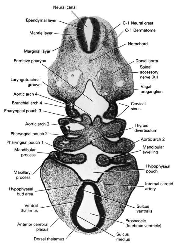
A section through the C-1 neural crest and the middle of the branchial arches and cervical sinus.
Observe:
1. The close relation of the hypophyseal bud area to the hypophyseal pouch.
2. The maxillary and mandibular processes of the first branchial arch.
3. Pharyngeal pouches 1 through 3 and aortic arches 2 through 4.
4. The point of origin of the thyroid diverticulum at the level of the second branchial arch and the laryngotracheal groove in the floor of the primitive pharynx.
5. The C-1 neural crest and its close relation to the dermatome.
Keywords: C-1 neural crest, anterior cerebral plexus, aortic arch 2, aortic arch 3, aortic arch 4, c-1 dermatome, cervical sinus, dorsal aorta, dorsal thalamus, ependymal layer, hypophyseal pouch, hypophyseal bud area, internal carotid artery, laryngotracheal groove, mandibular prominence of pharyngeal arch 1, mandibular swelling, mantle layer, marginal layer, maxillary prominence of pharyngeal arch 1, neural canal, notochord, pharyngeal arch 4, pharyngeal pouch 1, pharyngeal pouch 2, pharyngeal pouch 3, primitive pharynx, prosocoele (forebrain ventricle), sulcus medius, sulcus ventralis, thyroid diverticulum, vagal preganglion (CN X), ventral thalamus
Source: Atlas of Human Embryos.