raymond f. gasser
ATLAS OF HUMAN EMBRYOS
Copyright © 1975 RF Gasser, PhD. All rights reserved. Reproduced with permission.Chapter V (Pages 47-102)
- External Appearance
- Nervous System
- Alimentary and Respiratory Systems
- Urogenital System
- Coelomic Cavity and Mesenteries
- Cardiovascular System
- Skeletal, Muscular and Integumentary Systems
- Special Sense Organs
the fifth week of life
early branchial arch and limb bud period
I. EXTERNAL APPEARANCE
Fig. 5–1AThe specimens measure approximately 4 to 8 mm in length and have up to 42 to 44 pairs of somites.
The configuration of the embryo changes greatly, bending ventrally into a C shape.
The lateral, head and tail body folds move the attachment of the amnion to the midventral area in contact with the umbilical cord. In this area the coelom communicates with the extraembryonic coelom through a narrow channel between the attachment of the amnion and the yolk sac.
Bulges of the ectoderm form prominences in the head region that indicate the position of several structures.
The rostral and caudal neuropores have closed and are no longer evident.
Up to four branchial arches and grooves become distinct in the future lower face and neck region. The stomodeum or primitive mouth is bound by the maxillary and mandibular processes of the first arch. The fourth arch is in the floor of a depressed area called the cervical sinus.
The heart is prominent and located ventrally between the branchial arches and the yolk sac. The atrial and ventricular chambers are large and can be distinguished through the ectoderm.
The mesonephros, umbilical veins and spinal cord form bulges in the ectoderm.
The upper and lower limb buds appear as blunt projections on the lateral surface. A blunt, fingerlike tail bud becomes prominent in the midline in the most caudal part of the embryo.
The cloacal membrane is located in the midline on the ventral side between the lower limb buds.
II. NERVOUS SYSTEM
A. CENTRAL NERVOUS SYSTEM
Fig. 5–1BGENERAL
The neural folds have fused in the midline throughout the length of the embryo forming the neural tube on the dorsal side from head to tail. The overlying ectoderm is adjacent to the tube.
The cranial portion of the neural tube begins to enlarge to form the brain leaving the long, caudal portion to become the spinal cord. The rostral wall of the tube, where the rostral neuropore closes, remains thin and constitutes the lamina terminalis.
The neural tube becomes C shaped with two sharp ventral bends, one in the midbrain area, the cephalic flexure, the other at the brain-spinal cord junction, the cervical flexure.
The layer of proliferating neuroepithelial cells around the lumen of the neural tube is called the ependymal layer. Peripheral to this layer, neuroblasts collect in a middle layer called the mantle layer. Neuroblastic processes begin to collect in the periphery of the tube producing a relatively clear zone called the marginal layer.
BRAIN
After the neural folds fuse, the brain is subdivided into three dilations or vesicles named the prosencephalon (forebrain), mesencephalon (midbrain) and rhombencephalon (hindbrain). The cranial end of the prosencephalon is called the lamina terminalis.
The lumen of each vesicle forms a ventricle called the prosocoele, mesocoele and rhombocoele, respectively. The roof of the rhombocoele is very thin.
The optic vesicle develops as an outpouching that allows for further subdividing of the prosencephalon into telencephalon and diencephalon. The optic vesicle is considered part of the diencephalon.
-
Shallow grooves appear in the lateral wall of the brain separating its subdivisions into various portions.
Diencephalic part of prosencephalon—The sulcus medius separates the region of the dorsal thalamus from the ventral thalamus and is continuous caudally with the sulcus limitans. The epithalamus develops in the roof, in front of the pretectum. The sulcus ventralis separates the ventral thalamus from the hypothalamus and is continuous rostrally into the optic stalk. That part of the hypothalamus that is adjacent to the hypophyseal pouch is the neurohypophyseal bud area. The more rostral part where the optic stalk attaches is the optic chiasmal area.
Mesencephalon—The sulcus limitans divides the wall longitudinally into a dorsal part, the tectum, and a ventral part, the tegmentum. The basis pedunculi area will form ventral to the tegmentum.
Rhombencephalon—A series of vertical grooves segment its ventrolateral wall into six rhombomeres (neuromeres). This segmentation is partially evident on the outside of the tube as well. In the caudal portion the sulcus limitans separates the rhombencephalon into dorsolateral (alar plate) and ventrolateral (basal plate) areas.
SPINAL CORD
The long, caudal segment of the neural tube becomes the spinal cord. It gradually tapers in the tail region where it terminates as the primitive filum terminale.
The wall is thin in the midline dorsally and ventrally forming roof and floor plates, respectively. These areas will serve as pathways for crossing nerve fibers. The sulcus limitans divides the mantle layer in the lateral wall into a) a dorsolateral portion called the alar plate in which sensory nuclei form and b) a ventrolateral portion called the basal plate in which motor nuclei form.
B. PERIPHERAL NERVOUS SYSTEM
Fig. 5–2ANeural crest cells become abundant in the angle between the dorsolateral wall of the neural tube and the surface ectoderm.
CRANIAL NERVES
Optic Nerve (II)
See Section VIII, A.
Cranial Neural Crests
The neural crest cells along the cranial part of the neural tube collect into columns that extend ventrolaterally to become closely applied to the epibranchial placodes.
The cranial neural crests are more organized initially than the spinal neural crests (see below).
Experimental studies (Johnston MC: A radioautographic study of the migration and fate of cranial neural crest cells in the chick embryo. Anat Rec 156:143–156, 1966) indicate that cranial neural crest cells in birds are precursors of a) sensory ganglion cells of cranial nerves, b) autonomic (motor) ganglion cells on cranial nerves, c) neurolemmal sheaths of cranial nerves and d) branchial arch cartilages.
-
One neural crest is associated with each of the four branchial arches:
First branchial arch → trigeminal crest → cranial nerve V
Second branchial arch (including otocyst) → facioacoustic crest → cranial nerves VII and VIII
Third branchial arch → glossopharyngeal crest → cranial nerve IX
Fourth branchial arch → vagal crest → cranial nerve X
-
Sensory preganglia begin to form within each crest near the placode and will become ganglia on the cranial nerves.
Trigeminal crest → trigeminal ganglion (V)
Facioacoustic crest → geniculate (VII), cochlear (spiral) and vestibular (VIII) ganglia
Glossopharyngeal crest → superior (jugular) and inferior (petrous) ganglia (IX)
Vagal crest → superior (jugular) and inferior (nodose) ganglia (X)

FIG. 5–1 [printable version of FIG 5-1]
- External features of the 5-mm embryo.
- Central nervous system of the 5-mm embryo. Arrows indicate the position of the cephalic and cervical flexures.
Accessory Nerve (XI)
The accessory nerve is subdivided into cranial and spinal parts. The cranial part is a caudal extension of the vagal crest and is considered a part of the vagus nerve.
The spinal part first appears as a longitudinal collection of cells along the dorsolateral surface of the rhombencephalon and upper spinal cord. Rostrally it joins with the cranial part before it proceeds along the dorsal side of the precardinal vein. It disappears in a mesodermal condensation just caudal to the cervical sinus where the sternocleidomastoid-trapezius premuscle mass will form.
Hypoglossal Nerve (XII)
The hypoglossal nerve begins as a short, poorly defined, longitudinal collection of cells along the ventrolateral surface of the rhombencephalon. This cell collection is closely related to a condensation of occipital myoblasts.
SPINAL NERVES
Spinal Neural Crests
The crest cells along the spinal part of the neural tube collect together in the form of a longitudinal band on either side of the tube. The band has a scalloped ventral border. It is broadest in the upper cervical region and tapers toward the tail region.
Collections of crest cells extend farthest ventrolaterally near the middle of each body segment. Such extensions occur in a regular manner and are most prominent in the upper cervical segments.
A spinal ganglion (sensory) begins to develop within each crest midway between the neural tube and the myotome.
In some segments, crest cells have begun to spread ventral to the neural tube around the dorsal aorta. They will form the ganglia of the autonomic nervous system and are motor to the viscera.
The upper limb bud forms at the level of the C-5 to T-1 neural crests. The lower limb bud forms at the level of the L-1 to S-2 neural crests.
III. ALIMENTARY AND RESPIRATORY SYSTEMS
Fig. 5–2BA. ORONASOFACIAL REGION
Fig. 5–1AAn ectodermal depression called the stomodeum develops in the ventral part of the head region in the center of the developing face.
The mandibular process of the first branchial arch joins its counterpart on the other side in the midline to form the floor or caudal wall of the stomodeum. The two processes are separated on the surface by the median mandibular groove.
The maxillary process of the first branchial arch makes up the side or lateral wall of the stomodeum and is separated from the mandibular process by the primitive oral fissure.
The roof or rostral wall contains an evagination, the hypophyseal or Rathke’s pouch. The ectodermal lining of the pouch is adjacent to the neurohypophyseal area of the hypothalamus. An ectodermal thickening called the olfactory placode develops in the roof on each side, lateral to the telencephalic part of the prosencephalon.
B. FOREGUT
The oropharyngeal membrane ruptures and quickly disappears. As a result the endodermal lining of the foregut becomes continuous with the ectodermal lining of the stomodeum.
Five pharyngeal pouches appear on each side as blind, lateral extensions of foregut endoderm. This part of the foregut forms the primitive pharynx. Each of the first four pouches comes into contact with the ectoderm lining its corresponding branchial groove on the outside. The area of contact or closing membrane separates the pouch from the groove. The fifth pouch is actually a caudal extension of the fourth and is called the ultimobranchial body.
The point of attachment of the midline thyroid diverticulum moves slightly caudally to the level of the second aortic arch. The diverticulum becomes a small mass of cells adjacent to the aortic sac.
Respiratory system—The laryngotracheal groove deepens, converting the midportion of the foregut into a narrow tracheoesophageal tube that is compressed in the vertical plane. Behind the heart the tube separates into a small, dorsal primitive esophagus and a large, ventral primitive trachea. After a very short caudal course the primitive trachea separates into right and left lung buds.
The primitive esophagus is the caudal continuation of the foregut and terminates in a dilated segment of the endodermal tube, the primitive stomach.
Just caudal to the primitive stomach a small, dorsal midline diverticulum, the dorsal pancreatic bud, becomes evident.

FIG. 5–2 [printable version of FIG 5-2]
- Peripheral nervous system of the 5-mm embryo. Preganglion cells (large stipple) are evident in the cranial neural crests and form near placodes (striped areas).
- Alimentary and respiratory systems of the 5-mm embryo.
C. MIDGUT
A ventral, midline outpouching, the hepatic diverticulum, appears at the fore- and midgut junction, or primitive duodenum. The proximal part of the diverticulum dilates into the hepatic antrum, which communicates with the lumen of the primitive duodenum. Hepatic trabeculae, or cords of cells, extend from the antrum on both sides into the mesoderm of the septum transversum. The trabeculae are separated from each other by venous channels called hepatic sinusoids. The trabeculae join with one another between sinusoids.
A small outpouching, the ventral pancreatic bud, extends caudally from the hepatic antrum.
Between the hepatic antrum and the yolk stalk the midgut elongates in the vertical plane. The midportion of the midgut communicates with the yolk sac ventrally through the narrow yolk stalk. Caudal to the yolk stalk the midgut endoderm takes the shape of a small, cylindrical tube with a barely visible lumen. It is surrounded by the splanchnic layer of lateral mesoderm and is suspended in the coelom by a mesentery through which blood vessels reach the gut.
Because there are no distinguishing characteristics at this time, the mid- and hindgut junction becomes an arbitrary point on the caudal segment of gut.
D. HINDGUT
The cranial part of the hindgut extends caudally, then ventrally to become continuous with the rectal portion of the cloaca. It also is suspended from the dorsal body wall by a mesentery.
The caudal part of the hindgut dilates into a large chamber called the cloaca. The cloacal endoderm is elongated in the dorsoventral plane and makes contact with ectoderm in the midline of the future external genitalia. The area of contact is called the cloacal membrane.
A midline mass of mesoderm called the urorectal septum begins to divide the cloaca into two parts: a) a ventral urogenital sinus and b) a dorsal rectum.
The hindgut ends blindly in the tail bud as the tailgut.
IV. UROGENITAL SYSTEM
Fig. 5–3A. PRONEPHROS
All remnants of the pronephros disappear.
B. MESONEPHROS
As the pronephros regresses, excretory vesicles and tubules of the second embryonic kidney, the mesonephros, develop in the intermediate mesoderm in the C-6 to L-2 segments.
One end of each excretory tubule is cup shaped and is known as the glomerular capsule. The glomerular capsule surrounds a tuft of vascular channels and together they compose a corpsule. Each tubule terminates dorsolaterally into a common longitudinal channel, the mesonephric (Wolffian) duct. The duct begins in the lower cervical segments and terminates caudally in the urogenital sinus part of the cloaca. The more primitive vesicles that, as yet, have no connections with the duct are located in the caudal part of the mesonephros.
The tubules and vesicles form a bulge in the dorsolateral part of the coelom called the mesonephric ridge.

FIG. 5–3 [printable version of FIG 5-3]
Urogenital system of the 5-mm embryo.
C. GONAD
The coelomic epithelium between the mesonephros and the dorsal mesentery thickens to form the gonadal ridge. The gonadal ridge is apparent in the C-7 to T-8 segments. The mesonephric and gonadal ridges are suspended from the dorsal body wall by a thick urogenital mesentery. The primordial germ cells are migrating from the wall of the yolk sac to the gonadal ridge. They must migrate past the gut and its mesentery before reaching the ridge.
D. UROGENITAL SINUS
The urogenital sinus part of the cloaca receives the mesonephric ducts and the allantois. The allantois extends as an open channel into the umbilical cord. Its blind distal segment begins to disappear.
V. COELOMIC CAVITY AND MESENTERIES
A. COELOMIC CAVITY
The communication between the coelomic cavity and the extraembryonic coelom is reduced to a narrow channel on each side of the yolk stalk.
Caudal to the yolk stalk the coelomic cavity is a common chamber ventral to the primitive gut. Cranially it separates into right and left portions that communicate with the pericardial cavity through the pericardioperitoneal canals. A lung bud bulges into the cranial part of each canal as the caudal part narrows because of the expanding hepatic trabeculae and common cardinal and hepatocardiac veins.
The two canals are separated in the midline ventrally by the septum transversum. The septum transversum is an oblique plate of dense cellular tissue between the heart and the hepatic trabeculae. It represents the primitive diaphragm. The coelomic cavity caudal to the septum transversum will become the peritoneal cavity.
Recess on each side of the mesonephric ridge are called medial and lateral coelomic bays.
The entire coelomic cavity is lined with a single layer of cells called mesothelium.
B. MESENTERIES
Dorsal to the primitive gut the mesothelial layer on each side approaches in the midline to form the dorsal mesentery.
The two mesothelial layers are separated by a thick layer of mesodermal cells in which blood and lymph vessels to the gut and their derivatives develop. Nerves also reach these structures through the mesentery. The splenic primordium originates from the mesodermal cells in the dorsal mesentery in the region of the primitive stomach. The mesentery is named according to the structure it suspends (e.g., mesoesophagus, mesogastrium and mesoduodenum).
The mesocardium migrates to a position ventral to the esophagus, trachea and lung buds.
The mesonephros and gonadal ridge become suspended from the dorsal body wall on each side of the midline by the broad urogenital mesentery.
VI. CARDIOVASCULAR SYSTEM
A. HEART
Figs. 5–4, 5–5The heart tube is located primarily in the upper cervical segments and remains suspended dorsally by the mesocardium. Continued growth of the tube moves the bulboventricular part in a ventrocaudal direction. The initially paired atria, which were outside the pericardial cavity, fuse into a common chamber and move in a dorsocranial direction into the pericardial cavity. As a result of both movements the atrioventricular junction comes to lie in a more central position. The heart remains a single tube, but the location of its four definitive chambers becomes apparent.
The wall of the primitive ventricle becomes trabeculated to form the primitive left ventricle.
-
The bulbus cordis lies on the ventral side to the right with the deep bulboventricular sulcus at its left margin. The bulbus cordis can be subdivided into three parts:
The proximal part next to the primitive left ventricle is large with trabeculated walls and forms the primitive right ventricle. The primitive right and left ventricles are separated on the outside by a shallow interventricular groove. On the inside they communicate through the large primary interventricular foramen that is bounded cranially by a ridge of myocardium called the bulboventricular flange.
The middle part is cone shaped and called the conus cordis. It has smooth walls and courses obliquely toward the midline to form the outflow channels of both ventricles.
The distal part is the narrow, smooth-walled truncus arteriosus. It is located in the midline and joins the aortic sac.
The common atrial chamber communicates with the primitive left ventricle through the single atrioventricular (A-V) canal near the middle of the heart.
The common atrial chamber is divided into right and left portions by a ventral depression caused by the conus cordis and truncus arteriosus. The portions communicate with each other through a constricted area called the ostium primum. As each portion of the atrial chamber bulges cranially, a ridge proliferation between them produces the septum primum. The atrial walls remain temporarily smooth. Muscular trabeculae begin to form in the walls and produce bulges on the inner surface. The bulges will develop into muscular ridges called musculi pectinati.

FIG. 5–4 [printable version of FIG 5-4]
Heart of the 5-mm embryo.
Initially, the paired primordia of the sinus venosus, like the atria, are located entirely outside the pericardial cavity. They fuse into a common chamber and move in a dorsocranial direction. The chamber is not yet entirely within the pericardial cavity, coming to lie between the cavity and the hepatic trabeculae. It develops a large right and a small left horn. The right horn dilates and joins with the right atrium through the large sinoatrial orifice. A redundant fold of the wall to the right of the orifice forms the right venous valve. Each sinus horn receives three veins: a) the common cardinal vein, b) the umbilical vein and c) the vitelline vein by way of the hepatocardiac vein.
The walls of the heart tube remain double layered with a thin inner layer called the endocardium and a thick outer layer called the epimyocardium. A trabeculum of muscle tissue forms in the epimyocardium in the region of the ventricles. The endomyocardial space continues to separate the two layers except in the sinoatrial region.

FIG. 5–5 [printable version of FIG 5-5]
Cardiovascular system of the 5-mm embryo.
- Arteries
- Veins
B. ARTERIES
Fig. 5–5AFour pairs of aortic arches become apparent and connect the aortic sac with the paired dorsal aortas above the primitive pharynx. The first pair loses its connection with the dorsal aorta and begins to disappear in the mandibular process of the first branchial arch. The internal carotid artery is the rostral continuation of the dorsal aorta and lies in the roof of the primitive pharynx on each side of the hypophyseal pouch. The caudal three pairs of aortic arches course through the central portion of their respective branchial arch.
The large, paired dorsal aortas course caudally on either side of the midline between the notochord and the primitive gut. They gradually approach each other and fuse in the midline in the midcervical region. In the thoracic and upper lumbar regions the dorsal aorta is a large, single vessel. It bifurcates into right and left common iliac arteries in the lower lumbar region. The median sacral artery continues in the midline into the tail bud. After each common iliac artery gives rise to a small axial artery to the lower limb bud, it continues into the umbilical cord as an umbilical artery carrying blood to the chorionic villi. The umbilical arteries initially fuse in the proximal part of the cord but later separate completely.
-
Branches of the single dorsal aorta are divided into three groups:
Dorsal intersegmental arteries, which arise at every level between somites to supply the dorsal body wall and the spinal cord. In the region of each limb bud, one artery gives off a lateral branch that enlarges to form the axial artery.
Lateral segmental arteries, which supply the derivatives of the intermediate mesoderm (pro- , meso- , and metanephroi).
Ventral segmental arteries, which course through the dorsal mesentery to supply the gut and its derivatives. The artery to the fore- and midgut junction area is the celiac artery. One of the arteries to the midgut proper will become the superior mesenteric artery and one to the hindgut will become the inferior mesenteric artery. Several of the arteries to the midgut join together ventral to the gut to form the large vitelline artery. The vitelline artery courses in the yolk stalk bringing blood to the vitelline plexus in the yolk sac wall.
C. VEINS
Fig. 5–5BThree distinct pairs of venous channels bring blood to the sinus venosus: 1) vitelline veins from the yolk sac, 2) umbilical veins from the chorionic villi and 3) cardinal veins from the embryo proper.
Vitelline veins—After draining the yolk sac the right and left vitelline veins form dorsal and ventral anastomoses around the primitive duodenum. Then they break up into hepatic sinusoids as a result of invasion by the hepatic trabeculae. The sinusoids drain into the horns of the sinus venosus by short right and left hepatocardiac veins.
Umbilical veins—Blood in the chorionic villi moves to the embryo through the common umbilical vein in the umbilical cord. As the vein enters the embryo it divides into right and left umbilical veins, which break up into an extensive plexus in the lateral body wall. Each vein reforms and courses in the lateral body wall to join its respective horn of the sinus venosus. The proximal part of the left umbilical vein begins to disappear and in the region of the hepatic trabeculae anastomoses with the hepatic sinusoids.
-
Cardinal veins—Blood from the cranial part of the embryo drains to the heart through right and left precardinal veins. Blood from the caudal part of the embryo drains to the heart through right and left postcardinal veins. The pre- and postcardinal veins join on each side of the heart to form a short common cardinal vein, which enters its respective horn of the sinus venosus.
Precardinal vein—The cranial segment of the precardinal vein is called the primary head vein, which receives the anterior, middle and posterior cerebral plexuses. It passes medial to the trigeminal ganglion but ventrolateral to the otocyst. The precardinal veins course to the heart lateral to the dorsal aortas.
Postcardinal vein—The postcardinal vein begins in the tail bud and passes in a longitudinal manner toward the heart in the lateral part of the dorsal body wall between the somites and mesonephros. It receives intersegmental veins at every level. A new longitudinal channel called the subcardinal vein begins to develop along the medial side of the mesonephros. It gradually enlarges and eventually takes over the drainage of the expanding mesonephros. The subcardinal vein drains into the postcardinal vein by way of transverse anastomoses.
VII. SKELETAL, MUSCULAR AND INTEGUMENTARY SYSTEMS
A. HEAD MESODERM
The extrinsic ocular muscles will form in the mesenchymal tissue dorsal to the optic cup. A series of muscles and supporting structures will differentiate in the mesodermal core of the branchial arches. The muscles of the tongue will develop in the mesenchyme in the floor of the pharynx.
B. SOMITIC (PARAXIAL) MESODERM
Up to 42 to 44 somites differentiate on each side of the neural tube from the rhombencephalon to the coccygeal or tail region. There are four occipital, eight cervical, twelve thoracic, five lumbar, five sacral and eight to ten coccygeal. The first occipital somite disappears. The last five to seven coccygeal somites will disappear.
Cells in the ventral and medial walls of the somites actively proliferate and migrate toward the neural tube and notochord to form the sclerotome in each segment. This activity begins in the more cranial somites and proceeds in a caudal direction. Each sclerotome is separated by a dorsal intersegmental artery.
Cells in the dorsal wall of the somite retain their epitheloid character a while longer and compose the dermatome that is adjacent to the single-layered ectoderm.
Lightly staining, spindle shaped cells called myoblasts collect along the medial side of the dermatome. These cells form a platelike structure in each segment called the myotome, which will give rise to skeletal muscles.
C. INTERMEDIATE MESODERM
From the intermediate mesoderm will come most of the components of the Urogenital System (see above).
D. LATERAL (PLATE) MESODERM
The lateral mesoderm is separated by the coelom into somatic and splanchnic layers each of which becomes more cellular.
The somatic layer contributes to the formation of the ventro-lateral body wall.
The splanchnic layer surrounds the primitive gut. Condensed, splanchnic mesoderm in the dorsal mesogastrium will form the splenic primordium.
E. LIMB MESODERM
The limb buds contain dense mesoderm surrounded by thickened ectoderm or placode. The bones and muscles of the extremities form within this mesoderm.
Various studies on lower forms suggest three possible origins for this tissue: a) somites, b) lateral mesoderm or c) placode.
The upper limb bud forms at the level of neural crests C-5 to T-1. The lower limb bud forms at the level of neural crests L-1 to S-2. These segments are retained in the definitive condition and correspond to the segments of the motor and sensory nerves of the extremities.
VIII. SPECIAL SENSE ORGANS
A. EYE
Figs. 5–1, 5–2With closure of the prosencephalic part of the neural tube, the optic groove evaginates to form the optic vesicle. The vesicle is attached to the rostral part of the diencephalon by the optic stalk. The lumen within the vesicle and stalk is continuous with the prosocoele.
The floor of the diencephalon where the optic stalks join is called the chiasmal part of the hypothalamus.
Ectoderm adjacent to the optic vesicle thickens to form the lens placode.
B. EAR
Fig. 5–2AINTERNAL EAR
Fig. 5–2A
Placode VIII (otic) invaginates to produce the hollow, egg shaped otocyst from which the membranous labyrinth of the internal ear develops.
The acoustic part (VIII) of the facioacoustic neural crest terminates on the rostral surface of the otocyst.
MIDDLE EAR
Fig. 5–2B
The dorsal part of the first pharyngeal pouch comes into contact with ectoderm lining the dorsal part of the first branchial groove. The area of contact is the closing membrane or primitive tympanic membrane.
The auditory tube is derived from the proximal part of the first pharyngeal pouch, whereas the distal (lateral) part becomes the primitive middle ear cavity.
EXTERNAL EAR
Fig. 5–1A
The external auditory meatus arises from the dorsal part of the first branchial groove.
The auricle develops from the tissue surrounding the groove in the first and second branchial arches.
C. OLFACTORY APPARATUS
THE 5-MM EMBRYO
STAGE 13
AGE 28 DAYS
CROWN TO RUMP LENGTH 5.3 MM (33 SOMITES)
Carnegie collection 8066
Reference
Streeter GL: Developmental horizons in human embryos. Descriptions of age groups XIII, embryos about 4 to 5 millimeters long, and age group XIV, period of indentation of the lens vesicle. Contrib Embryol Carnegie Instn 31:27–47, 1945
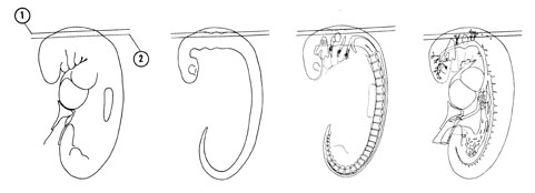
FIG. 5–6
SECTION 1
A section through the roof of the rhombencephalon.
Observe:
- The continuity between ectoderm and neural ectoderm where the rostral neuropore closed.
- The thin-walled roof of the rhombocoele (hindbrain ventricle), which will be invaginated by blood vessels to form a choroid plexus.
A section through the thicker, dorsal part of the rhombencephalon and the dorsal part of the otocyst.
Observe:
- The otocyst that will become the membranous labyrinth of the internal ear is still connected to the ectoderm.
- The close relation of the venous plexuses to the brain.

FIG. 5–6 [printable version of FIG 5-6]
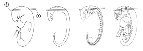
FIG. 5–7
SECTION 3
A section through the rhombencephalon, proximal part of the cranial neural crests and middle of the otocyst.
Observe:
- The pre- and postotic parts of the rhombencephalon in longitudinal section.
- The close relation of the otocyst to the rhombencephalon.
- Rhombomeres 1 through 6 in the wall of the rhombencephalon.
- The four cranial neural crests (two rostral to the otocyst and two caudal), which will form cranial nerves V, VII, VIII, IX and X.
- The close relation of the facioacoustic crest to the rostral side of the otocyst.
A section through the ventral part of the rhombencephalon and otocyst.
Observe:
- Rhombomeres 1 through 6 in the wall of the rhombencephalon.
- The two parts of the facioacoustic crest with the acoustic part terminating on the otocyst.
- The formation of the trigeminal preganglion adjacent to the ectoderm.
- The primary head vein that is lateral to the otocyst but medial to the trigeminal preganglion.
- The spinal accessory nerve between the rhombencephalon and occipital myotomes.

FIG. 5–7 [printable version of FIG 5-7]
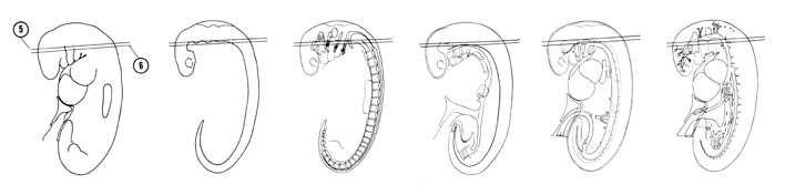
FIG. 5–8
SECTION 5
A section through the mesencephalon, rhombencephalon and dorsal part of the first branchial groove.
Observe:
- The ventral edge of the rhombencephalon in the midline.
- The caudal part of the rhombencephalon that will become the medulla oblongata.
- The mesencephalon in transverse section.
- The distal part of the four cranial neural crests (V, VII, IX and X) that enter the dorsal part of the branchial arches.
- The close relation of the vagal crest (X) to the lateral side of the primary head vein.
A section through the mesencephalon and rhombencephalon at the cephalic and cervical flexures, respectively.
Observe:
- The edge of the notochord and dorsal aortas.
- The junction of the geniculate preganglion with placode VII on the second branchial arch.
- The junction of the primary head vein with the precardinal vein.
- The occipital myotomes and their close association with the hypoglossal nerve that will innervate the tongue musculature.
- The dorsal part of branchial arches 1 and 2.

FIG. 5–8 [printable version of FIG 5-8]
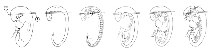
FIG. 5–9
SECTION 7
A section through the mesencephalon, rhombencephalon and cranial edge of the primitive pharynx.
Observe:
- The internal carotid artery, which is the rostral continuation of the dorsal aorta.
- The first and second branchial arches separated by the first branchial groove.
- The close association of the preganglia to the ectoderm.
- The close association of the glossopharyngeal preganglion to placode IX-X.
- The three parts of the definitive mesencephalon: tectum, tegmentum and basis pedunculi area.
A section through the junctions of the rhombencephalon with the spinal cord and the mesencephalon with the prosencephalon.
Observe:
- The branchial arches and the closing membranes separating the pouches from the grooves.
- The cranial end of the notochord at the hypophyseal pouch with the internal carotid arteries lateral.
- The junction of the large third aortic arch with the dorsal aorta.
- The close relation of the spinal accessory nerve to the precardinal vein.
- The paired dorsal aortas on each side of the primitive pharynx.

FIG. 5–9 [printable version of FIG 5-9]
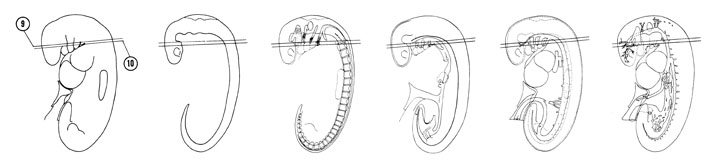
FIG. 5–10
SECTION 9
A section through the cranial edge of the C-1 neural crest and the floor of the primitive pharynx.
Observe:
- The edge of the hypothalamus and its relation to the hypophyseal pouch and internal carotid arteries.
- Branchial arches 1 through 4 with closing membranes separating pouches from grooves.
- The hypobranchial eminence in the midline in the floor of the primitive pharynx (tongue region).
- The close association of the vagal preganglion with placode IX-X.
- The junction of aortic arch 4 with the dorsal aorta and its close association to the primitive pharynx.
A section through the C-1 neural crest and the middle of the branchial arches and cervical sinus.
Observe:
- The close relation of the hypophyseal bud area to the hypophyseal pouch.
- The maxillary and mandibular processes of the first branchial arch.
- Pharyngeal pouches 1 through 3 and aortic arches 2 through 4.
- The point of origin of the thyroid diverticulum at the level of the second branchial arch and the laryngotracheal groove in the floor of the primitive pharynx.
- The C-1 neural crest and its close relation to the dermatome.

FIG. 5–10 [printable version of FIG 5-10]
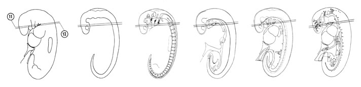
FIG. 5–11
SECTION 11
A section through the C-1–C-2 intercrest region, thyroid diverticulum and aortic sac.
Observe:
- The stomodeum separating the maxillary and mandibular processes.
- Pharyngeal pouches 1 through 4 and their relation to the aortic arches in the floor of the primitive pharynx.
- The origin of the aortic arches from the aortic sac.
- Myoblastic collection ventral to the C-1 myotome forming the sternocleido-mastoid-trapezius premuscle mass.
- The close relation of the C-1 neural crest to the C-1 myotome.
A section through the edge of the C-2 neural crest and the edge of the optic vesicle.
Observe:
- The close relation of the optic vesicle to the ectoderm and the maxillary process.
- The ventral part of the branchial arches and their close relation to the aortic sac.
- Pharyngeal pouch 5 (ultimobranchial body), which is a caudal continuation of pouch 4.
- The right dorsal aorta giving rise to a dorsal intersegmental artery.
- Sclerotome cells ventrolateral to the neural tube.

FIG. 5–11 [printable version of FIG 5-11]
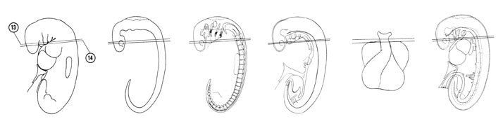
FIG. 5–12
SECTION 13
A section through the C-2 neural crest, truncus arteriosus and the ventral part of branchial arches 1 and 2.
Observe:
- The close relation between the prosencephalon and the overlying ectoderm.
- The optic stalk that will become the optic nerve connecting the optic vesicle to the diencephalic part of the prosencephalon.
- Branchial arches 1 and 2 filled with mesenchymal cells and the disappearance of the more caudal smaller arches.
- The caudal extent of the fifth pharyngeal pouch.
- The caudal part of the pharynx becoming a vertical slit where it is continuous with the tracheoesophageal tube in Section 14.
A section through the C-2 neural crest, middle of the optic vesicle and cranial edge of the heart.
Observe:
- The slightly thickened ectoderm (lens placode) adjacent to the optic vesicle.
- The communication between the prosocoele and the optic cavity.
- The median mandibular groove between the ventral end of the mandibular processes.
- The cranial edge of the pericardial cavity and the right and left atria separated by the septum primum.
- The cranial part of the tracheoesophageal tube where the larynx will develop.

FIG. 5–12 [printable version of FIG 5-12]
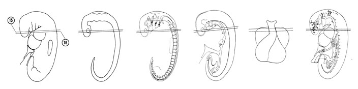
FIG. 5–13
SECTION 15
A section through the C-2 neural crest and the cranial edge of the ostium primum.
Observe:
- The relation of the mandibular process to the truncus arteriosus of the heart.
- The tracheoesophageal tube with its tracheal and espohageal parts.
- The close relation of the neural crest to the myotome.
- The location of the heart (atria) in the cervical region. Cardiac nerves originate in the cervical region and migrate with the heart into the thorax.
- The communication of the right and left atria through the ostium primum.
A section through the C-2–C-3 intercrest region and the junction of the telencephalic and diencephalic parts of the prosencephalon.
Observe:
- The edge of the optic stalk joining the prosencephalon.
- The inner endocardial layer of the conus cordis and the outer epimyocardial layer separated by the endomyocardial space.
- The mesocardium between the arterial (cranial) and venous (caudal) ends of the heart.
- The pericardial cavity lined with mesothelium.
- The dorsal position of the tracheoesophageal tube to the heart.

FIG. 5–13 [printable version of FIG 5-13]
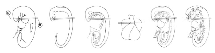
FIG. 5–14
SECTION 17
A section through the middle of the C-3 neural crest, telencephalic part of the prosencephalon and cranial part of the sinus venosus.
Observe:
- The dorsal position of the sinus venosus to the right atrium.
- The ventral position of the tracheal part of the tracheoesophageal tube to the esophageal part.
- The pre- and postcardinal veins near their junction to form the common cardinal vein.
A section through the C-3 neural crest and the separation of the tracheoesophageal tube.
Observe:
- The right venous valve between the sinus venosus and the right atrium.
- The bifurcation of the trachea into right and left lung buds that are primordia of the right and left bronchial trees.
- The lateral position of the pericardioperitoneal canal to the lung buds.
- The right and left common cardinal veins.
- The four plate regions of the neural tube in the C-3 segment.

FIG. 5–14 [printable version of FIG 5-14]
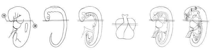
FIG. 5–15
SECTION 19
A section through the C-3 neural crest and the tracheal bifurcation.
Observe:
- The olfactory placodes located ventrolateral to the telencephalic part of the prosencephalon.
- The entrance of the right common cardinal vein into the sinus venosus.
- The right and left lung buds.
- The lateral position of the postcardinal veins to the paired dorsal aortas.
A section through the C-4 neural crest and the cranial part of the primitive left ventricle.
Observe:
- The cranial end of the prosencephalon called the lamina terminalis.
- The junction of the right hepatocardiac vein with the sinus venosus.
- The temporary fusion of the dorsal aortas.
- The origin of a dorsal intersegmental artery.

FIG. 5–15 [printable version of FIG 5-15]
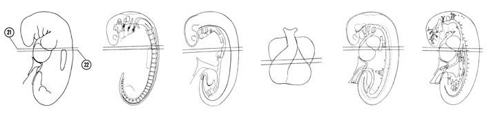
FIG. 5–16
SECTION 21
A section through the C-4 neural crest, upper limb bud, primitive stomach and the horns of the sinus venosus.
Observe:
- The continuity of the conus cordis in Section 20 with the primitive right ventricle of the heart.
- The communication (atrioventricular canal) between the atria and primitive left ventricle in Section 22.
- The junction of the right umbilical and hepatocardiac veins with the right sinus horn and the junction of the left common cardinal and umbilical veins with the left sinus horn.
- The slightly enlarged portion of the foregut that forms the primitive stomach.
- The trabeculae of the primitive left ventricle.
A section through the C-5 neural crest, cranial part of the hepatic trabeculae, septum transversum and upper limb bud.
Observe:
- The dorsal position of the sinus venosus to the primitive ventricles.
- The junction of the left hepatocardiac vein with the sinus venosus.
- The ventral mesogastrium between the stomach and hepatic trabeculae.
- The dorsal mesogastrium between the common dorsal aorta and the stomach.
- The common dorsal aorta at the level of the C-5 segment.

FIG. 5–16 [printable version of FIG 5-16]
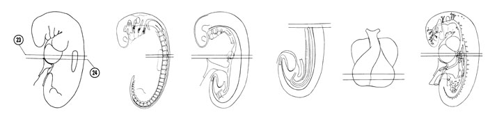
FIG. 5–17
SECTION 23
A section through the C-5 neural crest and the primary interventricular foramen.
Observe:
- The interventricular sulcus separating the primitive right and left ventricles and the caudalmost extent of the sinus venosus in the septum transversum.
- The hepatic trabeculae invading the septum transversum.
- The anastomosis of the vitelline veins ventral to the primitive duodenum and the umbilical veins in the lateral body wall.
- The dorsal pancreatic bud from which the body and tail of the pancreas develop.
- The communication of the hepatocardiac veins with the venous channels between the hepatic trabeculae.
A section through the C-6 neural crest, cranial end of the mesonephros and hepatic antrum.
Observe:
- The attachment of the amnion to the ventral body wall.
- The origin of the hepatic antrum from the ventral aspect of the duodenum. The antrum will give rise to the gall bladder and bile ducts.
- The cranial end of the vitelline veins.
- The medial position of the subcardinal vein to the mesonephros.
- The cranial part of the yolk sac.

FIG. 5–17 [printable version of FIG 5-17]
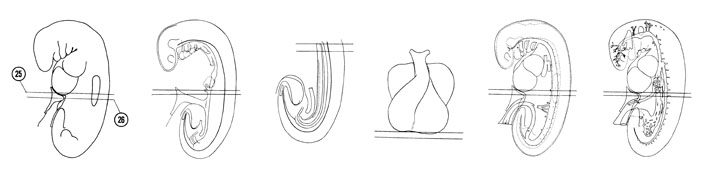
FIG. 5–18
SECTION 25
A section through the C-5 neural crest and the primary interventricular foramen.
Observe:
- The interventricular sulcus separating the primitive right and left ventricles and the caudalmost extent of the sinus venosus in the septum transversum.
- The hepatic trabeculae invading the septum transversum.
- The anastomosis of the vitelline veins ventral to the primitive duodenum and the umbilical veins in the lateral body wall.
- The dorsal pancreatic bud from which the body and tail of the pancreas develop.
- The communication of the hepatocardiac veins with the venous channels between the hepatic trabeculae.
A section through the C-6 neural crest, cranial end of the mesonephros and hepatic antrum.
Observe:
- The attachment of the amnion to the ventral body wall.
- The origin of the hepatic antrum from the ventral aspect of the duodenum. The antrum will give rise to the gall bladder and bile ducts.
- The cranial end of the vitelline veins.
- The medial position of the subcardinal vein to the mesonephros.
- The cranial part of the yolk sac.

FIG. 5–18 [printable version of FIG 5-18]
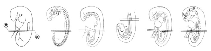
FIG. 5–19
SECTION 27
A section through the T-1 neural crest, the yolk stalk and the cranial edge of the umbilical cord.
Observe:
- The yolk stalk connecting the midgut with the yolk sac.
- The origin of a ventral segmental artery (superior mesenteric) from the dorsal aorta.
- The somatic and splanchnic parts of lateral mesoderm.
- The mesothelial lining of the coelom.
- The three layers of the neural tube.
A section through the T-1 neural crest and the middle of the midgut and yolk stalk junction.
Observe:
- The junction of the right and left umbilical arteries.
- The cranial edge of the allantois.
- The attachment of the amnion to the ventral body wall.
- The vitelline artery that is a continuation of one of the ventral segmental arteries.
- A mesonephric tubule joining its glomerular capsule with the mesonephric duct.

FIG. 5–19 [printable version of FIG 5-19]
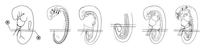
FIG. 5–20
SECTION 29
A section through the T-1–T-2 intercrest region and the caudal part of the midgut and yolk stalk junction.
Observe:
- The right and left branches of the common umbilical artery in the ventral body wall with the allantois between them.
- The course of the vitelline artery.
- The subdivisions of the left coelom into medial and lateral coelomic bays.
- The dorsal position of the postcardinal vein to the mesonephros.
- The plate regions of the neural tube in the thoracic region.
A section through the T-2 neural crest and the tip of the tail bud.
Observe:
- The tail bud containing the primitive filum terminale of the spinal cord, the caudal ends of the notochord and the tip of the tailgut.
- The common umbilical artery in the umbilical cord and the edge of the common umbilical vein.
- The common coelom which is isolated from the extraembryonic coelom.
- The midgut just caudal to the yolk stalk attachment.
- The origin of a dorsal intersegmental and a lateral and ventral segmental artery.

FIG. 5–20 [printable version of FIG 5-20]
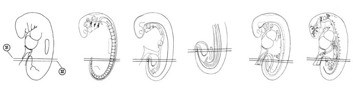
FIG. 5–21
SECTION 31
A section through the T-2–T-3 intercrest region, the caudal tip of the notochord and the mid- and hindgut junction
Observe:
- The lumen of the tailgut.
- The close association of the notochord with the primitive filum terminale dorsally and the tailgut ventrally.
- The attachment of the amnion to the ventral body wall at the umbilical cord.
- The larger size of the right umbilical vein compared to the left umbilical vein.
- A ventral segmental artery.
A section through the T-3 neural crest and the cranial part of the hindgut.
Observe:
- The caudal edge of the neural canal.
- The three primitive germ layers in the tail bud with their relative positions.
- Two subdivisions of lateral mesoderm: somatic and splanchnic.
- The bifurcation of the common umbilical vein into right and left umbilical veins.
- The cranial part of the hindgut suspended by the dorsal mesentery.

FIG. 5–21 [printable version of FIG 5-21]
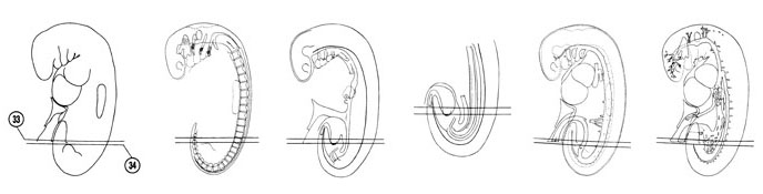
FIG. 5–22
SECTION 33
A section through the T-4–T-5 intercrest region and the cloacal membrane at the level of the S-3 somite.
Observe:
- The separation of the umbilical cord from the ventral body wall and the attachment of the amnion.
- The large vessels in the umbilical cord and the common umbilical artery separating into right and left branches.
- The S-3 somite and its relation to the neural tube.
- The cloacal membrane that is composed of ectoderm and endoderm and separates the cloaca from the exterior.
- The junction of the allantois with the urogenital sinus.
A section through the T-5–T-6 intercrest region and the junction of the mesonephric ducts with the cloaca at the level of the S-2 somite.
Observe:
- The median sacral artery (caudal continuation of the dorsal aorta) and its position between the notochord and the cloaca.
- The umbilical plexus of veins in the lateral body wall.
- The urogenital sinus between the umbilical arteries.
- The pelvic part of the coelom.
- The ventral segmental artery (inferior mesenteric) that courses from the dorsal aorta to the hindgut.

FIG. 5–22 [printable version of FIG 5-22]
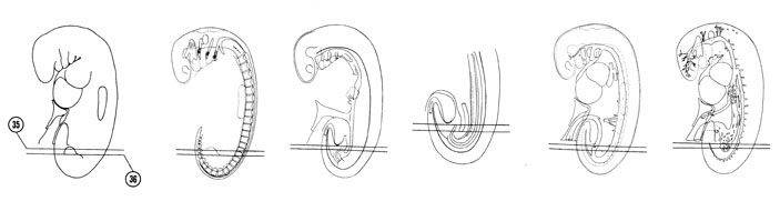
FIG. 5–23
SECTION 35
A section through the T-6 neural crest, S-1 somite and the cloaca.
Observe:
- The mycocoele of the S-1 somite surrounded by epitheloid cells.
- The urogenital sinus and rectal portions of the cloaca.
- The distal part of the lower limb buds with its ectodermal placode and mesodermal core.
- The caudal extent of the left umbilical plexus in the lateral body wall.
- The midsection of a mesonephric vesicle.
A section through the T-7 neural crest, L-5 somite and the urorectal septum.
Observe:
- The proximal part of the lower buds with the origin of the right axial artery from the common iliac artery.
- The junction of the umbilical and common iliac arteries.
- The mesodermal urorectal septum separating the hindgut from the cloaca.
- The left mesonephric duct in two locations.
- Somatic mesoderm in the abdominal wall.

FIG. 5–23 [printable version of FIG 5-23]
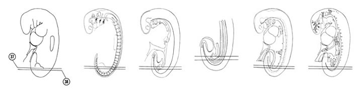
FIG. 5–24
SECTION 37
A section through the T-7 neural crest, L-5 somite and junction of the hindgut with the rectal portion of the cloaca.
Observe:
- The common iliac arteries lateral to the hindgut and cloaca junction.
- The proximal part of the lower limb bud.
- The caudal extent of the right umbilical plexus in the lateral body wall.
- The two segments of the mesonephric duct approaching each other.
A section through the T-8 neural crest, L-4 somite and aortic bifurcation.
Observe:
- The caudal extent of the postcardinal vein.
- The caudal end of the mesonephros (L-4 somite).
- The origin of the right and left common iliac arteries at the bifurcation of the dorsal aorta.
- The caudal extent of the gonadal ridge just medial to the mesonephros.
- The sclerotome cells between the notochord and the T-8 myotome.

FIG. 5–24 [printable version of FIG 5-24]
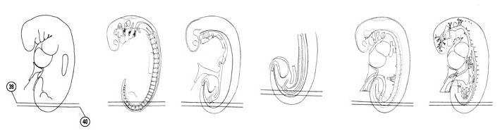
FIG. 5–25
SECTION 39
A section through the T-8 neural crest, L-3 dermatome and caudal part of the coelom.
Observe:
- Dermatomes in cross section (L-3) and oblique section (T-9).
- The near junction of the mesonephric ducts.
- The mesonephric vesicles and duct between the caudal part of the coelom and the dorsal aorta.
- The mesothelial lining of the coelom.
A section through the T-9–L-3 dermatomes and the adjacent body wall.
Observe:
- The caudal edge of the coelom on the right side.
- The dorsal edge of the dorsal aorta.
- The dorsal intersegmental arteries arising from the dorsal aorta.
- The T-10 to L-2 sclerotomes.

FIG. 5–25 [printable version of FIG 5-25]
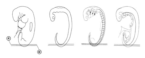
FIG. 5–26
SECTION 41
A section through the T-10–L-2 dermatomes and a longitudinal section of the notochord.
Observe:
- The close relation of the dermatomes to the ectoderm.
- The sclerotomes on each side of the notochord separated from one another by dorsal intersegmental arteries.
A longitudinal section of the basal plate of the spinal cord at the level of the T-11–L-2 dermatomes.
Observe:
- The neural crest cells between the plates of the spinal cord and the dermatomes.
- The neural canal in longitudinal section.

FIG. 5–26 [printable version of FIG 5-26]