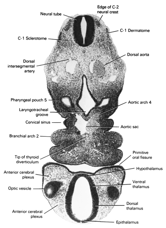
A section through the edge of the C-2 neural crest and the edge of the optic vesicle.
Observe:
1. The close relation of the optic vesicle to the ectoderm and the maxillary process.
2. The ventral part of the branchial arches and their close relation to the aortic sac.
3. Pharyngeal pouch 5 (ultimobranchial body), which is a caudal continuation of pouch 4.
4. The right dorsal aorta giving rise to a dorsal intersegmental artery.
5. Sclerotome cells ventrolateral to the neural tube.
Keywords: C-1 sclerotome, anterior cerebral plexus, aortic arch 4, aortic sac, c-1 dermatome, cervical sinus, dorsal aorta, dorsal intersegmental artery, dorsal thalamus, edge of C-2 neural crest, epithalamus, hypothalamus, laryngotracheal groove, neural tube, optic vesicle, pharyngeal arch 2, pharyngeal pouch 5, primitive oral fissure, tip of thyroid diverticulum, ventral thalamus
Source: Atlas of Human Embryos.