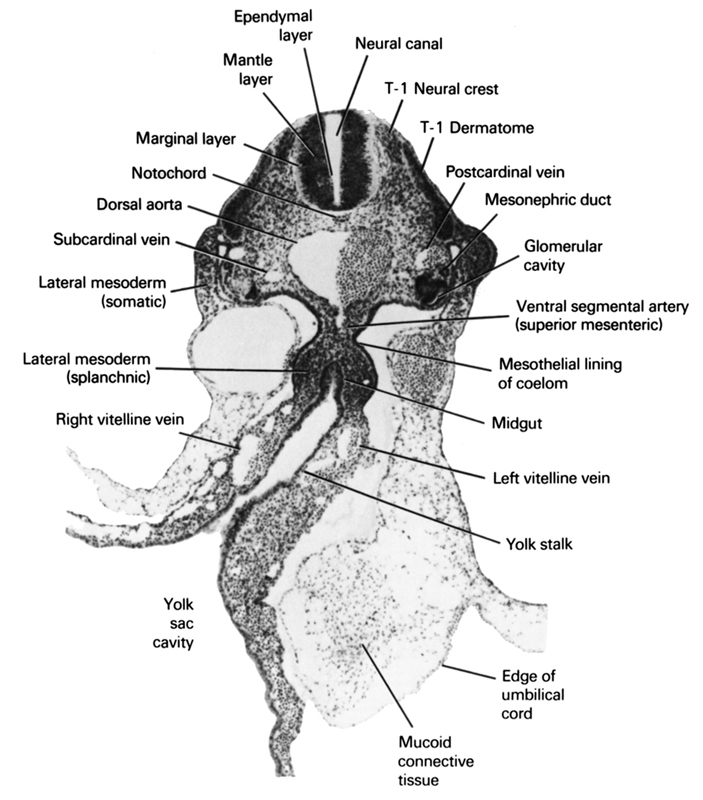
A section through the T-1 neural crest, the yolk stalk and the cranial edge of the umbilical cord.
Observe:
1. The yolk stalk connecting the midgut with the yolk sac.
2. The origin of a ventral segmental artery (superior mesenteric) from the dorsal aorta.
3. The somatic and splanchnic parts of lateral mesoderm.
4. The mesothelial lining of the coelom.
5. The three layers of the neural tube.
Keywords: T-1 dermatome, T-1 neural crest, dorsal aorta, edge of umbilical cord, ependymal layer, glomerular cavity, lateral mesoderm (somatic), lateral mesoderm (splanchnic), left vitelline (omphalomesenteric) vein, mantle layer, marginal layer, mesonephric duct, mesothelial lining of coelom, midgut, mucoid connective tissue, neural canal, notochord, postcardinal vein, right vitelline (omphalomesenteric) vein, subcardinal vein, ventral segmental artery (superior mesenteric), yolk sac, yolk sac cavity
Source: Atlas of Human Embryos.