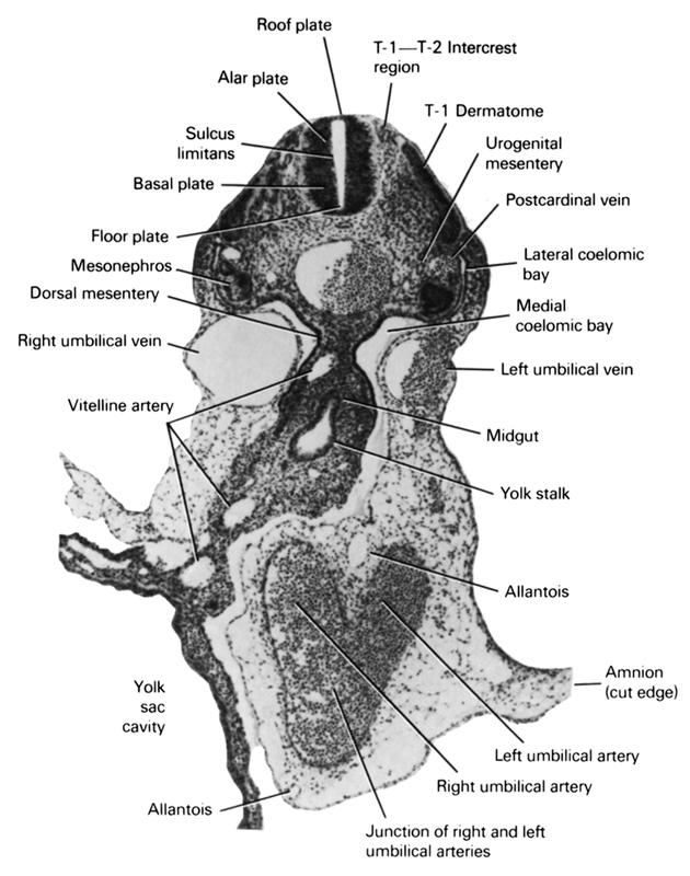
A section through the T-1–T-2 intercrest region and the caudal part of the midgut and yolk stalk junction.
Observe:
1. The right and left branches of the common umbilical artery in the ventral body wall with the allantois between them.
2. The course of the vitelline artery.
3. The subdivisions of the left coelom into medial and lateral coelomic bays.
4. The dorsal position of the postcardinal vein to the mesonephros.
5. The plate regions of the neural tube in the thoracic region.
Keywords: T-1 - T-2 intercrest region, T-1 dermatome, alar plate, allantois, amnion (cut edge), basal plate, dorsal mesentery, floor plate, junction of right and left umbilical arteries, lateral coelomic bay, left umbilical artery, left umbilical vein, medial coelomic bay, mesonephros, midgut, postcardinal vein, right umbilical artery, right umbilical vein, roof plate, sulcus limitans, urogenital mesentery, vitelline (omphalomesenteric) artery, yolk sac, yolk sac cavity
Source: Atlas of Human Embryos.