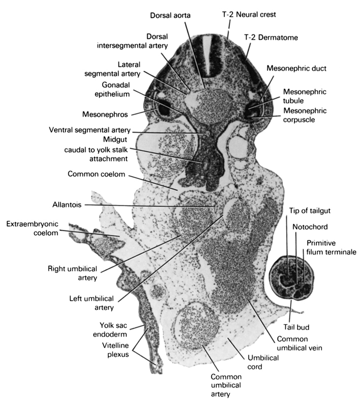
A section through the T-2 neural crest and the tip of the tail bud.
Observe:
1. The tail bud containing the primitive filum terminale of the spinal cord, the caudal ends of the notochord and the tip of the tailgut.
2. The common umbilical artery in the umbilical cord and the edge of the common umbilical vein.
3. The common coelom which is isolated from the extraembryonic coelom.
4. The midgut just caudal to the yolk stalk attachment.
5. The origin of a dorsal intersegmental and a lateral and ventral segmental artery.
Keywords: Midgut caudal to yolk sac attachment, T-2 dermatome, T-2 neural crest, allantois, common coelom, common umbilical artery, common umbilical vein, dorsal aorta, dorsal intersegmental artery, extraembryonic coelom, gonadal epithelium, lateral segmental artery, left umbilical artery, mesonephric corpuscle, mesonephric duct, mesonephric tubule, mesonephros, notochord, primitive filum terminale, right umbilical artery, tail bud, tip of tailgut, umbilical cord, ventral segmental artery, vitelline plexus, yolk sac endoderm
Source: Atlas of Human Embryos.