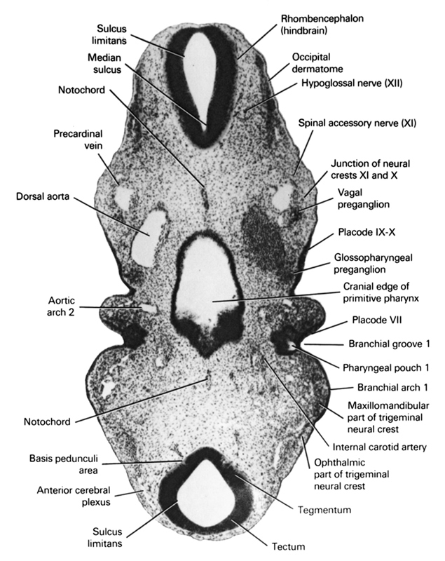
A section through the mesencephalon, rhombencephalon and cranial edge of the primitive pharynx.
Observe:
1. The internal carotid artery, which is the rostral continuation of the dorsal aorta.
2. The first and second branchial arches separated by the first branchial groove.
3. The close association of the preganglia to the ectoderm.
4. The close association of the glossopharyngeal preganglion to placode IX-X.
5. The three parts of the definitive mesencephalon: tectum, tegmentum and basis pedunculi area.
Keywords: Cranial edge of primitive pharynx, anterior cerebral plexus, aortic arch 2, basis pedunculi area, dorsal aorta, glossopharyngeal preganglion (CN IX), hypoglossal nerve (XII), internal carotid artery, junction of neural crests XI and X, maxillomandibular part of trigeminal neural crest, median sulcus, notochord, occipital dermatome, ophthalmic part of trigeminal neural crest, pharyngeal arch 1, pharyngeal groove 1, pharyngeal pouch 1, placode 7, placode 9-10, precardinal vein, rhombencephalon (hindbrain), sulcus limitans, tectum, tegmentum, vagal preganglion (CN X)
Source: Atlas of Human Embryos.