Biochemical Brain Abnormality Found in School-Age Children Prenatally Exposed to Cocaine
Download PDF Version What is PDF?
Robert Mathias
Robert Mathias is a Staff Writer for NIDA NOTES.
Source: NIDA NOTES, Vol. 16, No. 4, October, 2001
Public Domain
Table of Contents (TOC)
Article: Biochemical Brain Abnormality Found in School-Age Children Prenatally Exposed to CocaineReferences


Exposure to
cocaine before birth may affect the way a child’s brain functions
many years later, according to a recent NIDA-funded study. The brain-imaging
study found a chemical abnormality in the brains of 8-year-old children
that may reflect alterations in metabolic processes that enable brain
cells to use energy and function properly, the researchers say.
 “These children were exposed to cocaine only during gestation and their
brains have had 8 years to recover from that exposure,” says Dr. Joseph
Frascella of NIDA’s Division of Treatment Research and Development. “It
is surprising that they are still showing these deficits so many years later.” The
new finding suggests that early exposure to drugs has more long-lasting effects
on the brain than previously thought, he notes.
“These children were exposed to cocaine only during gestation and their
brains have had 8 years to recover from that exposure,” says Dr. Joseph
Frascella of NIDA’s Division of Treatment Research and Development. “It
is surprising that they are still showing these deficits so many years later.” The
new finding suggests that early exposure to drugs has more long-lasting effects
on the brain than previously thought, he notes.
The nature and extent of possible developmental damage to infants and children
from prenatal exposure to cocaine has been the subject of much apprehension and
scientific study. In the 1980s, anecdotal reports of abnormalities among cocaine-exposed
children contributed to fears that these children were irreparably damaged and
would never be able to function in society. Subsequent scientific research has
dispelled such exaggerated concerns for the vast majority of prenatally exposed
children. NIDA-funded studies that have been tracking the development of groups
of cocaine-exposed babies through adolescence now indicate that most seem to
function normally, but some may have subtle impairments in their ability to control
emotions and focus attention that could put them at risk of behavioral and learning
difficulties.
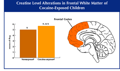
MRS scans suggest cocaine-exposed children did not have significant nerve damage or loss in the brain regions that were examined. However, cocaine-exposed children had significantly higher levels of the brain metabolite creatine than nonexposed children in a frontal area of the brain made up of "white matter," which consists mainly of nerve fibers and specialized support cells. The abnormality may reflect alterations in metabolic processes that enable brain cells to use energy and function properly.
Previous brain-imaging
studies of children prenatally exposed to cocaine have yielded conflicting
information about the drug’s effects on the developing central
nervous system. Some studies have found abnormalities in brain structure,
while others have not. Studies in abstinent adult cocaine abusers,
using an imaging technique called magnetic resonance spectroscopy
(MRS), have suggested that chronic cocaine use may cause persistent
damage to neurons in the frontal lobes of males and that brain metabolic
abnormalities also could exist despite a normal-appearing brain structure.
Dr. Lynne Smith of the Harbor-UCLA Medical Center in Torrance, California,
and Dr. Linda Chang of Brookhaven National Laboratory, in Upton, New
York, used this MRS technique to see if similar biochemical abnormalities
might be present in the brains of children who had been prenatally
exposed to cocaine, even if they appeared to have no structural damage.
The researchers used magnetic resonance imaging (MRI) to assess brain structure
and MRS to examine brain biochemistry in 14 8-year-old children who had been
exposed to cocaine in the womb. They administered the same brain scans to a control
group of 12 age-matched, nonexposed children. The MRS scans measured levels of
various chemicals in different brain regions. Increased or reduced concentrations
of these chemicals can indicate either damage to nerve cells or alterations in
brain cell function in these regions. The researchers assessed a frontal area
of the brain, made up of “white matter,” which consists mainly of
nerve fibers and specialized support cells. They also looked at an area deep
in the brain called the basal ganglia, which contains clusters of nerve cell
bodies, or “gray matter.”
 The study found no difference between the exposed and nonexposed children in
concentrations of N-acetyl-aspartate (NAA), a nerve cell metabolite, in either
the frontal area or the basal ganglia. Because NAA levels are markers for the
density and integrity of nerve cells, the normal NAA found in children prenatally
exposed to cocaine suggests they did not have significant nerve damage or loss
in the two brain regions that were examined. The MRI evaluations also showed
no brain structure abnormalities in children in either group. However, cocaine-exposed
children had significantly higher levels of creatine in the white matter of the
frontal lobes than nonexposed children. Elevated creatine levels indicate that
the brain cells of cocaine-exposed children use energy differently in this region.
The study found no difference between the exposed and nonexposed children in
concentrations of N-acetyl-aspartate (NAA), a nerve cell metabolite, in either
the frontal area or the basal ganglia. Because NAA levels are markers for the
density and integrity of nerve cells, the normal NAA found in children prenatally
exposed to cocaine suggests they did not have significant nerve damage or loss
in the two brain regions that were examined. The MRI evaluations also showed
no brain structure abnormalities in children in either group. However, cocaine-exposed
children had significantly higher levels of creatine in the white matter of the
frontal lobes than nonexposed children. Elevated creatine levels indicate that
the brain cells of cocaine-exposed children use energy differently in this region.
“All brain cells require creatine for all functions,” says Dr. Chang. “The
altered creatine levels we found could affect how both nerve cells and support
cells are functioning in the brain. We also have found the same abnormal creatine
levels in frontal white matter in adult cocaine abusers more than a year after
they have stopped using cocaine. The drug seems to have a particularly long-lasting
effect on energy metabolism in this brain area that merits further investigation.”
“The frontal area of the brain is involved in our ability to control impulses
and sustain attention on a task,” notes Dr. Frascella. Thus, it is possible
that the altered brain function found in this area could be a biological basis
for findings from other research that some cocaine-exposed children are more
impulsive and easily distracted than their peers. However, additional research
is needed to make this determination, he says.
Chang, L.; Ernst, T.; Strickland, T.; and Mehringer, C.M. Gender effects on persistent cerebral metabolic changes in the frontal lobes of abstinent cocaine users. American Journal of Psychiatry 156(5):716-722, 1999.
Chang, L., et al. Neurochemical alterations in asymptomatic abstinent cocaine users: A proton magnetic resonance spectroscopy study. Biological Psychiatry 42(12):1105-1114, 1997.
Smith, L.M.; Chang, L.; et al. Brain proton magnetic resonance spectroscopy and imaging in children exposed to cocaine in utero. Pediatrics 107(2):227-231, 2001.