836-2
The mass, together with the surrounding decidua and the underlying muscularis was carefully excised and placed in warm salt solution. Under a binocular microscope, the central portion of its free wall was carefully opened by a cruciate incision, which exposed the intervillus space. Through this was introduced a warm mixture of a saturated aqueous solution of mercuric chloride containing 5 per cent of glacial acetic acid, and the entire specimen was "fixed" by immersion in 500 cc. of the same solution. Afterwards it was hardened in alcohol, which was gradually increased from 50 to 70 percent.
On turning back the flaps of the cruciate incision, the intervillus space was exposed and found to be almost completely filled with villi. These averaged about 2.5 mm. in length, and each presented one or two main stems and a number of smaller bulbous branches. Under the dissecting microscope the chorionic membrane was then incised with a sharp scalpel, and the extra-embryonic coelom exposed. This measured 9 x 4 mm. and was in great part occupied by the embryo enclosed in its amnion, which was attached to the basal portion of the chorionic membrane. The umbilical vesicle measured 2 mm. in diameter and on its surface presented many irregularly shaped vessels and a uniform pattern of small blood vessels. The remainder of the coelomic cavity was traversed by strands of reticular magma, extending between the amnion and umbilical vesicle, as well as between them and the interior of the chorionic membrane.
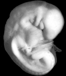
Shimmering through the amnion the 3 mm. embryo could be seen with its head visible above. It was apparently perfectly preserved and two gill arches were seen. The embryo was reconstructed by Dr. H. G. Evans, while the rest of the embryo forms the basis of this study.
Keywords: amnion, chorionic membrane, coelomic cavity, decidua, extra-embryonic coelom, intervillus space(s), muscularis, umbilical vesicle
Source: The Virtual Human Embryo.
