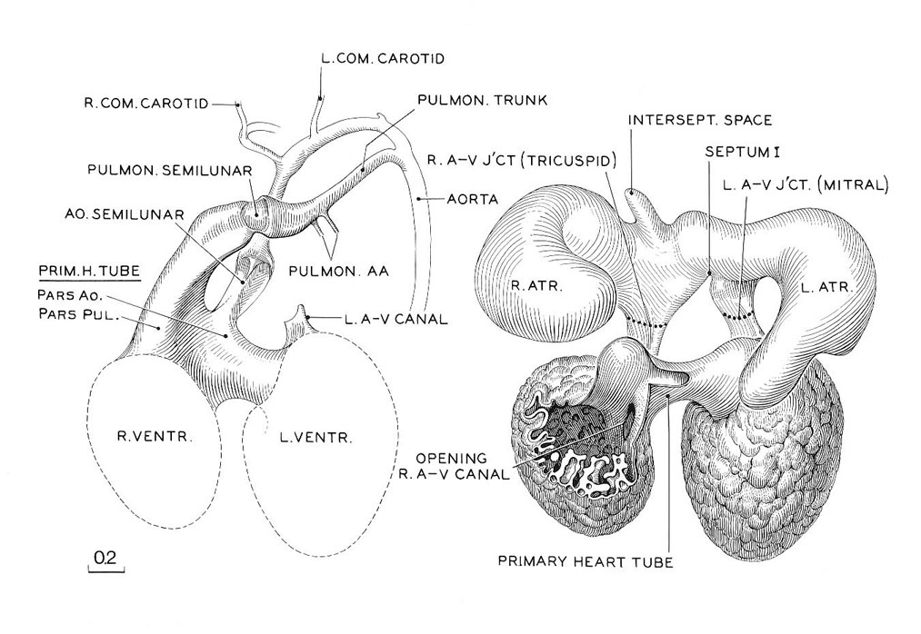
Reconstruction of the endocardium of the heart and its arterial trunks in No. 6520, a member of the median third of stage 17. It is shown in two parts, A and B. In A, only the derivatives of the primary cardiac tube are shown. In B, enough of the primary tube is removed to expose the venous part of the heart. The latter, being in a contracted state, reveals in an unobstructed view the right and left atrioventricular canals, now clearly separated, each leading forward into its respective ventricle. It will be noted that the primary cardiac tube is connected with the venous part of the heart only at the two atrioventricular junctions. When the latter are severed, the arterial portion can be freely separated from the venous part. In A, the cleavage has not yet crossed the crest of the septum, and both arterial trunks lead off from both ventricles. From the distention of the endocardium of the primary cardiac tube, however, one can see that the left ventricle favors the aorta and the right ventricle the pulmonary trunk.
O'Rahilly and Müller, 1987 Fig. 17-7
Keywords: aorta, aortic semilunar valve, arterial trunk, atrioventricular junction, endocardium, interseptal space, left atrioventricular (mitral) valve, left atrioventricular canal, left atrium, left common carotid artery, left ventricle, opening of right atrioventricular canal, pars aortic, pars pulmonic, primary cardiac tube, pulmonary artery, pulmonary semilunar valve, pulmonary trunk, right atrioventricular (tricuspid) valve, right atrioventricular canal, right atrium, right common carotid artery, right ventricle
Source: The Virtual Human Embryo.