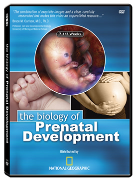
Multilingual Illustrated DVD [Tutorial]
The Biology of Prenatal Development
Introducing the Multilingual Illustrated DVD
Explore the fascinating imagery and facts presented in The Biology of Prenatal Development at your own pace. Each clip from the program is accompanied by its corresponding written script. Select Play Movie to watch any clip. Select See Snapshots to view high resolution images. See the program script and subtitles in 88 languages by using the Choose Language drop-down menu and clicking Refresh. Subtitles are displayed in your chosen language and may be turned on and off by clicking the ![]() button found in the lower right corner of the movie player. A "full screen" option is also available by clicking the
button found in the lower right corner of the movie player. A "full screen" option is also available by clicking the ![]() button.
button.
Download English PDF Download Spanish PDF Download French PDF What is PDF?
Table of Contents
- THE EMBRYONIC PERIOD (THE FIRST 8 WEEKS)
-
- EMBRYONIC DEVELOPMENT: THE FIRST 4 WEEKS
-
- Chapter 3 – Fertilization
- Chapter 4 – DNA, Cell Division, and Early Pregnancy Factor (EPF)
- Chapter 5 – Early Stages (Morula and Blastocyst) and Stem Cells
- Chapter 6 – 1 to 1 1/2 Weeks: Implantation and Human Chorionic Gonadotropin (hCG)
- Chapter 7 – The Placenta and Umbilical Cord
- Chapter 8 – Nutrition and Protection
- Chapter 9 – 2 to 4 Weeks: Germ Layers and Organ Formation
- Chapter 10 – 3 to 4 Weeks: The Folding of the Embryo
- EMBRYONIC DEVELOPMENT: 4 TO 6 WEEKS
-
- Chapter 11 – 4 Weeks: Amniotic Fluid
- Chapter 12 – The Heart in Action
- Chapter 13 – Brain Growth
- Chapter 14 – Limb Buds and Skin
- Chapter 15 – 5 Weeks: Cerebral Hemispheres
- Chapter 16 – Major Airways
- Chapter 17 – Liver and Kidneys
- Chapter 18 – Yolk Sac and Germ Cells
- Chapter 19 – Hand Plates and Cartilage
- EMBRYONIC DEVELOPMENT: 6 TO 8 WEEKS
-
- Chapter 20 – 6 Weeks: Motion and Sensation
- Chapter 21 – The External Ear and Blood Cell Formation
- Chapter 22 – The Diaphragm and Intestines
- Chapter 23 – Hand Plates and Brainwaves
- Chapter 24 – Nipple Formation
- Chapter 25 – Limb Development
- Chapter 26 – 7 Weeks: Hiccups and Startle Response
- Chapter 27 – The Maturing Heart
- Chapter 28 – Ovaries and Eyes
- Chapter 29 – Fingers and Toes
- THE 8-WEEK EMBRYO
- THE FETAL PERIOD (8 WEEKS THROUGH BIRTH)
-
- Chapter 37 – 9 Weeks: Swallows, Sighs, and Stretches
- Chapter 38 – 10 Weeks: Rolls Eyes and Yawns, Fingernails & Fingerprints
- Chapter 39 – 11 Weeks: Absorbs Glucose and Water
- Chapter 40 – 3 to 4 Months (12 to 16 Weeks): Taste Buds, Jaw Motion, Rooting Reflex, Quickening
- Chapter 41 – 4 to 5 Months (16 to 20 Weeks): Stress Response, Vernix Caseosa, Circadian Rhythms
- Chapter 42 – 5 to 6 Months (20 to 24 Weeks): Responds to Sound; Hair and Skin; Age of Viability
- Chapter 43 – 6 to 7 Months (24 to 28 Weeks): Blink-Startle; Pupils Respond to Light; Smell and Taste
- Chapter 44 – 7 to 8 Months (28 to 32 Weeks): Sound Discrimination, Behavioral States
- Chapter 45 – 8 to 9 Months (32 to 36 Weeks): Alveoli Formation, Firm Grasp, Taste Preferences
- Chapter 46 – 9 Months to Birth (36 Weeks through Birth)
The Embryonic Period (The First 8 Weeks)
Embryonic Development: The First 4 Weeks
Chapter 3 Fertilization
Biologically speaking,
"human development
begins at fertilization,"
when a woman and a man
each combine 23
of their own chromosomes
through the union
of their reproductive cells.
A woman's reproductive cell is commonly called an "egg" but the correct term is oocyte.
Likewise, a man's reproductive cell is widely known as a "sperm" but the preferred term is spermatozoon.
Following the release of an oocyte from a woman's ovary in a process called ovulation, the oocyte and spermatozoon join within one of the uterine tubes, which are often referred to as Fallopian tubes.
The uterine tubes link a woman's ovaries to her uterus or womb.
The resulting single-celled embryo is called a zygote, meaning "yoked or joined together."
Chapter 4 DNA, Cell Division, and Early Pregnancy Factor (EPF)
The zygote's 46 chromosomes
represent the unique
first edition
of a new individual's
complete genetic blueprint.
This master plan resides
in tightly coiled
molecules called DNA.
They contain the instructions
for the development
of the entire body.
DNA molecules resemble a twisted ladder known as a double helix. The rungs of the ladder are made up of paired molecules, or bases, called guanine, cytosine, adenine, and thymine.
DNA molecules resemble a twisted ladder known as a double helix. The rungs of the ladder are made up of paired molecules, or bases, called guanine, cytosine, adenine, and thymine.
Guanine pairs
only with cytosine,
and adenine with thymine.
Each human cell contains
approximately 3 billion
base pairs.
The DNA of a single cell contains so much information that if it were represented in printed words, simply listing the first letter of each base would require over 1.5 million pages of text!
If laid end-to-end, the DNA in a single human cell measures 3 1/3 feet or 1 meter.
The DNA of a single cell contains so much information that if it were represented in printed words, simply listing the first letter of each base would require over 1.5 million pages of text!
If laid end-to-end, the DNA in a single human cell measures 3 1/3 feet or 1 meter.
If we could uncoil
all of the DNA
within an adult's
100 trillion cells,
it would extend
over 63 billion miles.
This distance reaches from
the earth to the sun and back
340 times.
Approximately 24 to 30
hours after fertilization,
the zygote completes
its first cell division.
Through the process
of mitosis,
one cell splits into two,
two into four, and so on.
As early as 24 to 48 hours
after fertilization begins,
pregnancy can be confirmed
by detecting a hormone
called "early pregnancy factor"
in the mother's blood.
Chapter 5 Early Stages (Morula and Blastocyst) and Stem Cells
By 3 to 4 days
after fertilization,
the dividing cells of the embryo
assume a spherical shape
and the embryo is called
a morula.
By 4 to 5 days, a cavity forms within this ball of cells and the embryo is then called a blastocyst.
The cells inside the blastocyst are called the inner cell mass and give rise to the head, body, and other structures vital to the developing human.
Cells within the inner cell mass are called embryonic stem cells because they have the ability to form each of the more than 200 cell types contained in the human body.
Chapter 6 1 to 1½ Weeks: Implantation and Human Chorionic Gonadotropin (hCG)
After traveling
down the uterine tube,
the early embryo
embeds itself
into the inner wall
of the mother's uterus.
This process, called
implantation, begins 6 days
and ends 10 to 12
days after fertilization.
Cells from the growing embryo begin to produce a hormone called human chorionic gonadotropin, or hCG, the substance detected by most pregnancy tests.
HCG directs maternal hormones to interrupt the normal menstrual cycle, allowing pregnancy to continue.
Cells from the growing embryo begin to produce a hormone called human chorionic gonadotropin, or hCG, the substance detected by most pregnancy tests.
HCG directs maternal hormones to interrupt the normal menstrual cycle, allowing pregnancy to continue.
Chapter 7 The Placenta and Umbilical Cord
Following implantation,
cells on the periphery
of the blastocyst
give rise to part of
a structure called the placenta,
which serves as an interface
between the maternal
and embryonic
circulatory systems.
The placenta delivers maternal oxygen, nutrients, hormones, and medications to the developing human; removes all waste products; and prevents maternal blood from mixing with the blood of the embryo and fetus.
The placenta also produces hormones and maintains embryonic and fetal body temperature slightly above that of the mother's.
The placenta communicates with the developing human through the vessels of the umbilical cord.
The life support capabilities of the placenta rival those of intensive care units found in modern hospitals.
Chapter 8 Nutrition and Protection
By 1 week,
cells of the inner cell mass
form two layers called
the hypoblast
and epiblast.
The hypoblast gives rise to the yolk sac, which is one of the structures through which the mother supplies nutrients to the early embryo.
Cells from the epiblast form a membrane called the amnion, within which the embryo and later the fetus develop until birth.
The hypoblast gives rise to the yolk sac, which is one of the structures through which the mother supplies nutrients to the early embryo.
Cells from the epiblast form a membrane called the amnion, within which the embryo and later the fetus develop until birth.
Chapter 9 2 to 4 Weeks: Germ Layers and Organ Formation
By approximately 2 1/2 weeks,
the epiblast has formed
3 specialized tissues,
or germ layers,
called ectoderm,
endoderm,
and mesoderm.
Ectoderm gives rise to numerous structures including the brain, spinal cord, nerves, skin, nails, and hair.
Endoderm produces the lining of the respiratory system and digestive tract, and generates portions of major organs such as the liver and pancreas.
Mesoderm forms the heart, kidneys, bones, cartilage, muscles, blood cells, and other structures.
By 3 weeks
the brain is dividing
into 3 primary sections
called the forebrain,
midbrain,
and hindbrain.
Development of the respiratory and digestive systems is also underway.
Development of the respiratory and digestive systems is also underway.
As the first blood cells
appear in the yolk sac,
blood vessels form
throughout the embryo,
and the tubular heart emerges.
Almost immediately, the rapidly growing heart folds in upon itself as separate chambers begin to develop.
The heart begins beating 3 weeks and 1 day following fertilization.
The circulatory system is the first body system, or group of related organs, to achieve a functional state.
Almost immediately, the rapidly growing heart folds in upon itself as separate chambers begin to develop.
The heart begins beating 3 weeks and 1 day following fertilization.
The circulatory system is the first body system, or group of related organs, to achieve a functional state.
Chapter 10 3 to 4 Weeks: The Folding of the Embryo
Between 3 and 4 weeks,
the body plan emerges
as the brain,
spinal cord,
and heart of the embryo
are easily identified
alongside the yolk sac.
Rapid growth causes folding of the relatively flat embryo. This process incorporates part of the yolk sac into the lining of the digestive system and forms the chest and abdominal cavities of the developing human.
Rapid growth causes folding of the relatively flat embryo. This process incorporates part of the yolk sac into the lining of the digestive system and forms the chest and abdominal cavities of the developing human.