Skip Navigation

Prenatal Form and Function – The Making of an Earth Suit

  
[Help]

Unit 6:   5 to 6 Weeks

  Closer Look: 
  Applying the Science: 
Play Movie - Human Embryo 5 to 6 weeks.
Movie 6.1 - 5 to 6 Weeks
Brain and heart of a 5 week 6 day embyro.
Figure 6.1 - Five-Week Embryo (Note Brain & Heart)
The Biology of Prenatal Development DVD
Copyright © 2006 EHD, Inc. All rights reserved.


stressed out boy
Trying to comprehend the complexity of the network of synapses in the human brain is enough to make your head hurt!

Cartilage formation begins by 5½ weeks.1

By 5½ weeks, retinal pigment is subtly visible externally.2


By 6 weeks a portion of the brain called the cerebral cortex (ser’e-bral kor’teks) appears.3 Nerve cells, or neurons, in the spinal cord now begin to develop specialized connections.4  These connections, where neurons meet and communicate with one another, are called synapses.5

neuron
Figure 6.2 - Neuron
The neuron is perfectly designed to communicate with other nerve cells.
LifeART
Copyright © 2002 Lippincott, Williams & Wilkins.
Play Movie - The Rapidly Growing Brain
Movie 6.2 - The Rapidly Growing Brain


Movement Begins

Though a pregnant woman does not feel movement for at least another 8 to 10 weeks, the embryo begins to move between 5 and 6 weeks.6 The embryo’s first movements are both spontaneous and reflexive.7 A light touch to the mouth area causes the embryo to reflexively withdraw its head, 8 while the embryo’s trunk will twist spontaneously. Movements are essential for the normal development of bones and joints.9

 By 6 weeks, the external ear begins to take shape,10 and the opening of the ear canal becomes visible. Salivary glands also appear inside the mouth.11

Play Movie - Response to Touch
Movie 6.3 - Response to Touch
woman exercising and lifting weights
Just as weight lifting develops muscles through practice and repetitive motions, the embryo must “practice” movement to develop normal bones and joints.
Adult Ear
Figure 6.3 - Adult Ear
The adult ear is even more complex inside than outside. Amazingly the inner and outer ear begin forming just six weeks after fertilization.
LifeART
Copyright © 2002 Lippincott, Williams & Wilkins.
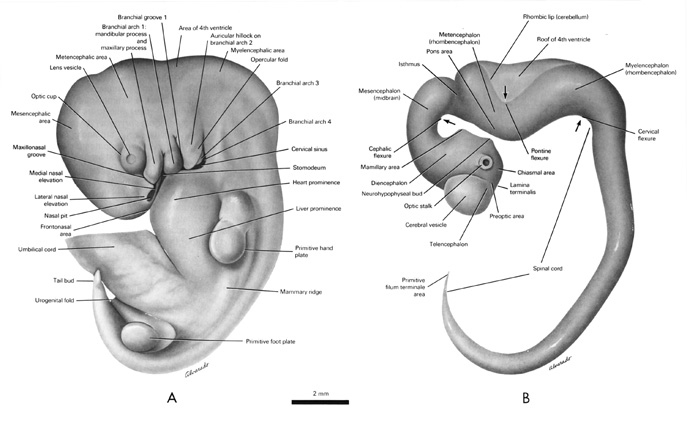

Open PDF version of FIG 6.4, External Features, Nervous System, of the 10mm Embryo
Figure 6.4 - External Features (A) and Nervous System (B) of the 10mm Embryo [PDF version of FIG 6.4]
From Gasser RF, 1975, 105. Atlas of Human Embryos. Copyright © 1975 RF Gasser, PhD. All rights reserved.


Blood formation is now actively underway in the liver12 and contributes to the liver’s bright red color.13 The rapidly growing liver also begins producing lymphocytes.14 This type of white blood cell is a key part of the developing immune system.

Play Movie - Blood Formation in Liver
Movie 6.4 - Blood Formation in Liver
Firefighter
Like a firefighter putting out a blazing fire, your immune system puts out fires in your body – stopping germs and infections from spreading and making you sick.


baby holding adults finger
Our hands begin to develop 5 weeks after fertilization.
Girl Playing with Blocks
 

By 5½ weeks, 5 linear digital rays begin forming the bones in the hand including the thumb and fingers.15 Wrist formation is also underway.16

At 6 weeks, the embryo’s hand plates develop a subtle flattening and linear digital rays now become noticeable.17 These digital rays form the bones of the fingers and metacarpals of each hand as development progresses.


By 5½ weeks, nipples appear near the embryo’s underarm.18 We will see their location change as they reach their final position on the chest wall.

The diaphragm (di’a-fram), the primary muscle used in breathing, is largely formed by 6 weeks.19

Diaphragm of 6 weeks 6 days embryo
Figure 6.5 - Diaphragm
The Biology of Prenatal Development DVD
Copyright © 2006 EHD, Inc. All rights reserved.
Play Movie - Nipple Formation
Movie 6.5 - Nipple Formation


By 6 weeks, a portion of the intestine begins to temporarily protrude outside the abdominal cavity. It extends into the umbilical cord. This normal process, called physiologic herniation (fiz-e-o-loj’ik her-ne-a’shun), makes room for other developing organs in the abdomen, such as the liver.20

Play Movie - Physiologic Herniation
Movie 6.6 - Physiologic Herniation
Physiologic herniation, intestine loops, appendix, and umbilical cord of the 7 week embryo.
Figure 6.6 - Physiologic Herniation
Notice the loops of intestine extending into the umbilical cord of this 7 week embryo. This is a normal and necessary part of embryonic development.
The Biology of Prenatal Development DVD
Copyright © 2006 EHD, Inc. All rights reserved.


Glass of milk
When we drink milk, the parathyroid hormone helps our body absorb calcium.
 

The endocrine system continues to develop during week 6, producing hormones which will help control numerous functions of the body. The parathyroid glands form in the neck area by 6 weeks and eventually secrete a hormone which helps regulate calcium levels in the blood.21 Over each kidney the adrenal cortex also forms and soon will secrete hormones which help manage stress and balance salt and fluid levels.

The pancreas now begins producing glucagon,22 an important hormone that prevents blood sugar levels from dropping dangerously low.

back to top
Take a written quiz about this unit. Take a visual quiz about this unit.
5 to 6
Weeks
Footnotes

1 Moore and Persaud, 2003. 382; O'Rahilly and Gardner, 1975. 4.
2 O'Rahilly and Müller, 2001. 459; O'Rahilly and Müller, 1987. 187.
3 Marin-Padilla, 1983. 21.
4 Cunningham FG et al., 2001. 149; de Vries et al., 1982. 320; Okado et al., 1979. 496.
5 Noback et al., 1996. 14.
6 Birnholz et al., 1978. 539; de Vries et al., 1982. 301, 304; Humphrey, 1970. 12; Kurjak and Kos, 1994. 48; Sorokin and Dierker, 1982. 723, 726; Visser et al., 1992. 175-176.
7 Birnholz et al., 1978. 539; de Vries et al., 1982. 301, 304; Hogg, 1941. 373; Humphrey, 1964. 99; Humphrey, 1970. 12; Humphrey and Hooker, 1961. 147; Humphrey and Hooker, 1959. 7; Humphrey and Hooker, 1959. 76; Kurjak and Kos, 1994. 48; Natsuyama, 1991. 13; O'Rahilly and Müller, 1999a. 336; Sorokin and Dierker, 1982. 723, 726; Visser et al., 1992. 175-176.
8 Goodlin, 1979. D-128; Humphrey, 1964. 99.
9 Humphrey, 1970. 35; Liley, 1986. 11; Reinis and Goldman, 1980. 224.
10 Karmody and Annino, 1995. 251; O'Rahilly and Müller, 2001. 480; Streeter, 1948. 190.
11 Grand et al., 1976. 793; Moore and Persaud, 2003. 220-221.
12 Moore and Persaud, 1993. 190.
13 Moore and Persaud, 2003. 262.
14 Kurjak and Kos, 1994. 19.
15 Moore and Persaud, 2003. 86.
16 Moore et al., 1994. Figure 2-16 A & B, 38-39.
17 O'Rahilly and Müller, 1987. 202-203.
18 Moore et al., 2000. 259; Moore and Persaud, 2003. 492; O'Rahilly and Müller, 1985. 155; Wells, 1954. 126.
19 de Vries et al., 1982. 320.
20 Gilbert-Barness and Debich-Spicer, 1997. 774; Grand et al., 1976. 798; Moore and Persaud, 2003. 267-268; O'Rahilly and Müller, 1987. 213; Sadler, 2005. 66; Spencer, 1960. 9; Timor-Tritsch et al., 1990. 287.
21 Moore and Persaud, 1998. 211.
22 Cunningham FG et al., 2001. 150.