Biologically speaking, fertilization (or conception) is the beginning of human development.1 Fertilization normally occurs within several hours of ovulation2 (some authors report up to 24 hours3) when a man’s sperm, or spermatozoon, combines with a woman’s egg, or secondary oocyte, inside a woman’s uterine tube (usually in the outer third of the uterine tube called the ampulla).4
Fertilization begins with the spermatozoon contacting the cells surrounding the oocyte and ends with the mixing of the 23 male and 23 female chromosomes.5 [More about fertilization] The result is a single-cell embryo called a zygote,6 meaning "yoked or joined together,"7 and it is the first cell of the human body.
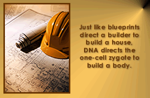
The zygote, like the oocyte, is encased by its protective covering, the zona pellucida, [More about the zona] 8 and contains 46 unique chromosomes with the entire genetic blueprint of a new individual. Chromosomes contain tightly packed, tightly coiled molecules called DNA.9 [More about DNA] Amazingly, DNA contains all the instructions needed for this single-cell embryo to develop into an adult.
The final steps in zygote formation include replication of the male and female DNA and the alignment of chromosomes in preparation for the first cell division through mitosis (mi-to’sis).10 The chromosomes assume a formation called a cleavage spindle, which is a phase of mitosis.
As the 2 sets of chromosomes migrate to opposite ends of the zygote, a crease begins to form along the equator marking the impending line of division.11 (Fig xx) The zygote or single-cell embryo completes the first cell division approximately 24 to 30 hours after fertilization.12 The process of repeated cell division is called cleavage.13
The process of fertilization joins the genetic contribution of a mother and father to form a complete set of DNA so that development can proceed. The structure of the spermatozoon and secondary oocyte (reviewed in the Introduction) are uniquely equipped for this process. To briefly summarize, the successful spermatozoon must combine speed (to reach and enter the oocyte first), strength (to help get through part of the corona radiata) and chemicals (to penetrate the corona radiata and the zona pellucida).14
Fertilization begins when the spermatozoon makes contact with the oocyte15 and involves several distinct steps:16
Penetration of the corona radiata
Penetration of the zona pellucida
The zona reaction
Fusion of male and female cell membranes
Formation of male and female pronuclei
Fusion of male and female pronuclei
1. Penetration of the Corona Radiata
Penetrating the surface cells of the oocyte (corona radiata) is accomplished in part through the spermatozoon’s "active swimming movements" 17 and in part through the use of digestive enzymes contained in the acrosome of the spermatozoon.
2. Penetration of the Zona Pellucida
Upon reaching the zona pellucida, the full dose of acrosomal enzymes is released. The process of enzyme release and hole formation in the zona is called the acrosomal reaction. This reaction also changes the spermatozoon membrane enabling it to bind to the oocyte membrane.18
3. The Zona Reaction
Entry of the first spermatozoon through the zona triggers a change throughout the entire zona pellucida which prevents additional spermatozoa from entering into the newly forming zygote. This "zona reaction" ensures that the DNA from only a single spermatozoon joins with the maternal DNA.19 The zona pellucida also helps prevent the embryo from implanting prematurely in the wall of the uterine tube - an example of a condition called ectopic pregnancy (More about ectopic pregnancy).20
4. Fusion of Male and Female Cell Membranes
The spermatozoon now travels quickly across the perivitelline space (a fluid filled space between the inside of the zona and the cell membrane of the oocyte) and attaches to the cell membrane of the oocyte. The cell membranes fuse forming one cell allowing free entry of the male nucleus (with tail attached) into the oocyte.
5. Formation of Male and Female Pronuclei
Fusion of the oocyte and spermatozoon cell membranes releases the oocyte to complete its second meiotic division. This process forms two unequal cells - a mature oocyte with almost all of the cell contents and a second polar body.21 (Sometimes, the first polar body also divides.)
The tightly packed nucleus of the mature oocyte now spreads out and forms the female pronucleus.
The tightly packed male nucleus loses its tail and also expands forming the male pronucleus.22
6. Fusion of Male and Female Pronuclei
Once the 2 pronuclei are formed, they enlarge, replicate their DNA in preparation for the first mitotic cell division,23 and draw close to one another. Their membranes degenerate allowing the female and male DNA to join for the first time. This marks the end of fertilization and this single cell is properly called a zygote.24
After the first cell division, these 2 daughter cells are called blastomeres and proceed with mitosis independently. Two cells become four, four become eight, and so on as these and subsequent daughter cells divide repeatedly.36 The first days of cell division do not increase the size of the embryo because the blastomeres become smaller as their numbers increase. Additionally, after the third round of cell division, the cells become more tightly packed in a process called compaction. As compaction proceeds, the cells of the embryo divide into 2 populations with distinct destinies. (described below)
Subsequent cell divisions occur take about 8 hours to reach completion as will become apparent by reviewing Figure 1.8 below. Replication of DNA over that time period requires an assembly rate exceeding 208,000 nucleotides per second.37Amazingly, DNA replication is accomplished with an average of only 1 error per billion (109) nucleotides.38 190?39
As early as 24 to 48 hours after fertilization begins, pregnancy can be confirmed by detecting a hormone called “early pregnancy factor” or EPF in the mother’s blood40 (The test for this hormone, however, is not widely available). This substance helps prevent the mother’s immune system from rejecting the soon-to-be-implanted embryo and allows pregnancy to proceed.41
Embryo Transport in the Uterine Tube
As cell division proceeds, the embryo is on the move. The uterine tube steers the embryo toward the uterus by cilia motion of epithelial cells lining the uterine tube wall and continued mild muscle contractions.42 The cells of the corona radiata disappear within about 2 days43 while the zona persists in protecting the embryo and preventing premature implantation in the uterine tube.
The ability of DNA to store such a mind-boggling quantity of information lies in its structure. This beautifully complex molecule resembles the shape of a twisted ladder, which is called a double helix.44 Alternating sugar and phosphate molecules link together to make up the sides of this ladder. The rungs of the ladder consist of molecules called bases: cytosine (si’to-sen), guanine (gwahn’en), adenine (a-den’en), and thymine (thi’men).45 Two of these molecules join to form a base pair, and connect one side of the ladder to the other by bonding to the sugar molecules. Cytosine always pairs with guanine and adenine with thymine. When bonded together, a sugar molecule, phosphate molecule, and base form a nucleotide, which is the building block of DNA.46 Each nucleotide is typically represented by the first letter of the base it contains: C, G, A, or T.47
The sequence of nucleotides within DNA forms a code or language. Every language has rules and researchers have discovered and carefully delineated the rules of this fascinating language. The "alphabet" of DNA has only four “letters” with each "letter" representing a nucleotide. All “words” are made of exactly three letters.48 This means there are a maximum of 64 words (4 × 4 × 4 = 64). Each word plays a crucial role in the process of protein synthesis.
Figure 1.4 - DNA's Double Helix
Courtesy: NHGRI - NIH.
Recent Advances
Reseachers who have recently mapped the human genome estimate human DNA contains between 30,000 and 40,000 genes,49 though most scientists previously believed that number to be much higher.50 These inherited units of information determine a person’s unique traits such as the color of our eyes, hair, and skin. As we grow and interact with our environment, genes also play a significant role in numerous other traits such as our eventual height, athletic ability, and susceptibility to genetic diseases.
The vast quantity of genetic information within the DNA of a single cell is overwhelming. We can begin to wrap our minds around this concept by using the “letters” of each base - C, G, A, and T. Each human cell contains approximately 3 billion ( 3 × 10 9) of these base pairs.51 Simply listing one letter for each base within a single cell would require over 1.5 million pages of text!52 If instead we unwind and stretch out the DNA in a single human cell, it will measure a full 3 1/3 feet or about 1 meter.53 Or we could stretch out the entire DNA from within an adult’s estimated 100 trillion (1 × 10¹⁴) cells, which end-to-end measures over 63 billion (6.3 × 10¹⁰) miles. This distance reaches from the earth, to the sun, and back 340 times.54
Figure 1.5 - Chromosomes
Courtesy: The National Human Genome Research Institute - National Institute of Health (NHGRI - NIH).
By about 3 days after fertilization, while still in the uterine tube, the embryo contains 12 to 16 cells configured as a solid ball of cells and is called a morula (mor’u-la) [Fig ss] 66
Approximately 3 1/2 to 4 days after fertilization, the uterine tube relaxes under the influence of progesterone67 and the embryo completes its journey through the uterine tube and enters the uterus.68 By this time the embryo begins to develop a fluid-filled cavity with a collection of cells at one end and is called a blastocyst.69 [Fig ee]
The surface of the blastocyst adjacent to the inner cell mass is referred to as the polar end or embryonic pole of the blastocyst.70 [Fig ee]
The zona pellucida, having delivered the embryo through the maze of the uterine tube, degenerates shortly after the embryo arrives in the uterus.71 Embryos studied outside the uterus in a process sometimes called “hatching.”72 The now-free blastocyst is now ready to find a permanent home inside the wall of the uterus.
The zona pellucida’s job is to protect and deliver viable embryos to the uterus and then disappear.
The zona pellucida, often referred to as the "zona," is an non-cellular glycoprotein that surrounds the oocyte and forms as each primary follicle develops from a primordial follicle within the ovary.73 It is formed jointly by granulosa cells surrounding the oocyte and the oocyte itself and contains the embryo for the first 4 to 5 days - enough time for the embryo to travel through the uterine tube and into the uterus.
The zona serves several vital functions. The zona protects the oocyte from entry by spermatozoa of a different species and from entry by more than one spermatozoon. The zona also triggers the acrosomal reaction necessary for successful fertilization and helps prevent premature implantation of the embryo in the uterine tube. 74
It should be noted that the zona pellucida is not a totally impermeable barrier (except to spermatozoa following the entry of the first spermatozoa). The oocyte and early embryo are made of living cells. These cells consume oxygen and nutrients which must be constantly available within the uterine tube, and they produce carbon dioxide which must be constantly removed. The nutrients produced and released by the cells lining the uterine tube are utilized by the embryo and therefore must be able to cross the zona.75 A truly impermeable barrier would be incompatible with survival.
So, why do we need the zona pellucida? We need the zona because without it, fertilization and transport through the uterine tube could not succeed. In summary, no zona, no embryo, no newborn!
Implantation is the process whereby the early embryo embeds into the inner wall of the mother’s uterus. Implantation begins about 6 days after fertilization and is complete by about 12 days.79
The first step of this process is the attachment phase, which begins about 6 days after fertilization80 [Fig rr]. The outer cells (trophoblast cells) of the blastocyst have specialized adhesion molecules81 which bind to the epithelial cells of the endometrium. The cells in the uterine wall are full of nutrients and water. The blastocyst attaches between the uterine glands, along its surface overlying the inner cell mass (the embryonic pole). Once attached. the trophoblast cells release enzymes that digest, liquify, and separate maternal cells forming an entry way inside the uterine wall. The trophoblast cells capture the local nutrients and actively share them with the inner cell mass. This will be discussed further in Unit 2.
Implantation represents a significant obstacle to the developing embryo. It is estimated that up to one-half of all embryos fail to successfully implant and die82 - often without the mother realizing she is pregnant. Many of these embryos are thought to have severe genetic abnormalities incompatible with survival.
By the end of the first week, the embryo has traveled extensively, multiplied from 1 cell to several hundred, dramatically changed its shape and complexity, and begun the process of finding permanent housing.
In Fig. 1.9 we see the embryo (at the blastocyst stage) embedding into the inner wall of the uterus in a process called implantation.
During the proliferative phase of the menstrual cycle (described in detail in the Introduction) estrogen is produced and released by the granulosa cells and theca interna cells of the developing follicles. Estrogen stimulates significant growth of the endometrium with proliferation of glands in the uterine wall accompanied by a rich blood supply.
Following ovulation and the onset of the secretory phase of the menstrual cycle, progesterone is released by the corpus luteum and further prepares the uterine wall for implantation. The blood supply continues to increase. Glycogen, a carbohydrate chain from which glucose is easily released, and other nutrients such as proteins, lipids, and minerals are produced and stored in the uterine glands.83 Water retention adds to the thickness of the uterine lining.84 Some of the water and nutrients are released into the uterine cavity to provide nourishment for the floating embryo prior to implantation.85 Approximately 6 days after fertilization, the lining of the uterus is maximally ready to begin implantation - just in time for the arrival of the embryo.
So, how does the uterus prepare for implantation? The uterus prepares by responding to various hormones levels to provide a protected environment with a rich blood supply, plenty of water, and abundant nutrients at just the right time..
Without a properly functioning uterus, the embryo would quickly perish!
Stem Cells — Unlimited Career Potential
From the first days of development, stem cells are present. These cells will differentiate into all of the 200 plus cell types contained in the human body!89 In fact, under the right conditions, an individual stem cell can separate from the embryo and form an entirely new embryo resulting in identical twins.
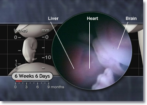
Figure 1.10 -The Embryonic Liver, Heart, and Brain
These cells are the building blocks from which every successive cell in the human body develops. Stem cells function as a reservoir of undifferentiated cells and are capable of dividing and becoming highly specialized cells. Stem cell research involves stimulating these cells in such a way as to cause them to develop into anything from a brain cell to a heart cell or a liver cell. This incredible capability of stem cells perfectly suits them for early prenatal development.
The specialized cells in our brain, heart, liver, and the rest of our body all originate from stem cells in the embryo.
Visual Summary of the First Week
Figure 1.11 below provides a visual summary of the events of the first week following fertilization. Note the zona pellucida surrounding the embryo through the 58-cell stage at 4 days but not at the 107-cell stage at 4½ days.
Figure 1.11 - The Many Forms and Locations of the Human Embryo During the First Week [PDF version of FIG 1.11]
Proteins carry out the instructions contained in DNA. There are tens of thousands of different proteins in our body including many hormones, clotting factors, enzymes, and antibodies for fighting infection. They also help give shape and stability to cell structure. Survival would be impossible without this wide variety of proteins.
The incredibly complex instructions in DNA, the intricate mechanism by which they are followed, and the impressive reliability of the whole process are almost beyond comprehension.
Proteins are a major class of macromolecules which make up many cellular components and which perform numerous vital functions. [See sidebar] The 20 amino acids in the human body are the building blocks of all proteins.90 Of the 64 words in this language of DNA, 60 represent a code for one of these 20 amino acids. Because the number of words (60) far exceeds the number of amino acids (20), there are synonyms – multiple words which encode the same amino acid. The remaining 4 words (of the original 64) signal the protein building process to either start or stop.91 The DNA code provides the instructions for building proteins or various kinds of ribonucleic acids or RNA. DNA ultimately controls the form and function and maintenance of each cell type in our bodies by controlling the types and quantities of proteins produced.
The Three Types of RNA1,2
I. Ribosomal RNA provides the workbench, combining with proteins to form ribosomes – the "workbench" on which proteins are synthesized. II. Transfer RNA delivers raw materials, binding specific amino acids and delivering them to the ribosome to be added to the protein under construction. III. Messenger RNA provides instructions, carrying the specific instruction code for each protein from DNA to the ribosome where the code is used to build each protein.
1 Lodish, Berk, Zipursky, et al., Molecular Cell Biology, 4th Ed. (New York: Freeman, 2000), 127.
2 Guyton and Hall, Textbook of Medical Physiology, 10th Ed. (Philadelphia: W. B. Saunders, 2000), 27.
Protein Synthesis – Courtesy of the RNA Trio
Protein synthesis is accomplished by “reading” these three-letter words and following their instructions. Each “word” dictates one particular amino acid and the order of the words dictates the sequence of amino acids needed for each protein. When a “stop” is encountered, the process ends. A section of DNA that encodes the complete instruction set for a protein molecule or an RNA molecule is called a gene.92
A full explanation of this complex process is well beyond the scope of this article but there are many excellent sources for the interested student.
The substantial effort put forth by the human body to capture and deliver the early embryo to the uterus can be better understood by looking at the challenges facing a woman whose embryo implants in an abnormal location.
For a variety of reasons (See Risk Factors below), the embryo may implant in the uterine tube which is by far the most common site of abnormal implantation. Implantation of the embryo in a site other than the body of the uterus is referred to as an "ectopic pregnancy" meaning "out of place" and represents a medical emergency.
Implantation of the embryo in the thin wall of the uterine tube can lead to rupture of the uterine tube accompanied by the sudden onset of heavy bleeding which can be life-threatening. Ectopic pregnancy is the leading cause of pregnancy-related death in the first trimester.
Risk Factors
Women most at risk for ectopic pregnancy include women with a history of previous ectopic pregnancy, pelvic inflammatory disease (PID) (especially when caused by Chlamydia trachomatis), tubal surgery (including tubal sterilization surgery), various uterine surgeries including assisted reproduction procedures, use of certain hormone medications, and hormone level abnormalities.
 Closer Look:
Fertilization |
DNA |
Zona Pellucida |
Uterus Preparation |
Proteins
Closer Look:
Fertilization |
DNA |
Zona Pellucida |
Uterus Preparation |
Proteins
 Applying the Science:
Ectopic Pregnancy
Applying the Science:
Ectopic Pregnancy


")