Prenatal Form and Function – The Making of an Earth Suit
Unit 8: 7 to 8 Weeks
 Closer Look:
 Applying the Science:

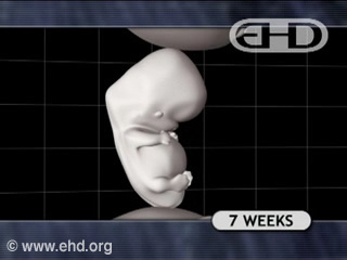
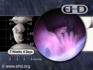
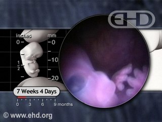
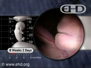
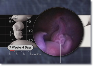
From 7 to 7½ weeks, tendons attach leg muscles to bones,1 and knee joints appear.2 Also by 7½ weeks, the hands can be brought together, as can the feet.3 The embryo also kicks, and will jump if startled.4
Also by 7 to 7½ weeks, nephrons, the basic filtration units in the kidneys, begin to form.
Copyright © 2006 EHD, Inc. All rights reserved.
Copyright © 2002 Lippincott, Williams & Wilkins.

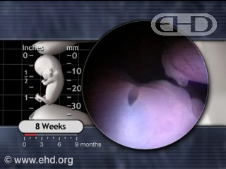
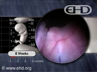
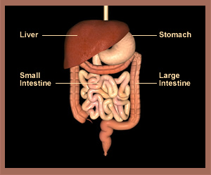
By 8 weeks peristalsis (per-i-stal’sis)5 begins in the embryo’s large intestine, a process which will continue throughout life. This alternating contraction and relaxation of the intestine wall is essential for proper digestion as it propels ingested food and fluids in the right direction through the intestine. Peristalsis also causes the release of gastrointestinal tract contents into the amniotic fluid in which the embryo floats.6The kidneys begin to produce urine, which is then released into the amniotic fluid.7
After swallowing our food, contractions of muscle within the intestine help push our food forward – an ability already developing in the 8-week embryo. Nephrons in the kidneys filter toxins out of the blood stream. Toxins then flow from the kidneys into the bladder and eventually out of the body. In the male embryo, developing testes begin to produce and release testosterone.8
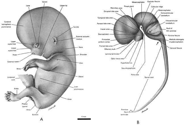
A Summary of Hand and Foot Development
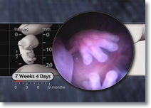
Copyright © 2006 EHD, Inc. All rights reserved.

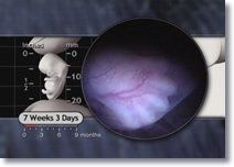
Copyright © 2006 EHD, Inc. All rights reserved.
Copyright © 2006 EHD, Inc. All rights reserved.

By 8 weeks the brain is highly developed9 and makes up approximately 43 percent of the embryo’s total weight.10 Growth continues at an extraordinary rate. One of the major control centers for the body - the hypothalamus - begins to take form. The hypothalamus eventually controls body temperature, heart rate, blood pressure, fluid balance, and the secretion of vitally important hormones by the pituitary gland.11
Our body’s temperature is regulated by the hypothalamus – an important structure which begins developing within the 8-week embryo’s brain.

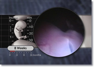
Slowly or rapidly, singularly or repetitively, spontaneously or reflexively, the embryo continues to practice the movements begun earlier and to move in new ways. Frequently, hands will touch the face and the head will turn.12 The many muscles of the face are now largely well developed in preparation for the complex facial expressions to follow.13 Touching the embryo can produce squinting, jaw movement, grasping motions, and toe pointing.14

Pediatric textbooks describe the ability to “roll over” as appearing between 10 and 20 weeks after birth for most infants.15 The fetus, however, displays this impressive coordination long before birth as shown in The Biology of Prenatal Development DVD. The low gravity environment of the fluid-filled amniotic sac16 resting in the uterus makes this maneuver possible. Only the lack of strength required to overcome the higher gravitational force outside the uterus prevents newborns from rolling over.17
Copyright © 2006 EHD, Inc. All rights reserved.
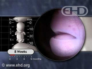
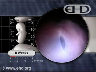
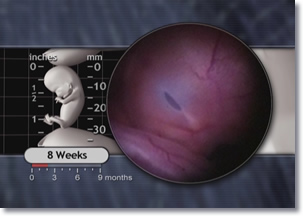
Between 7 and 8 weeks the upper and lower eyelids grow rapidly and begin to fuse together, giving the eyes a nearly closed appearance by 8 weeks.18 The eyelids are easily visible and by 7½ weeks are poised to enter a stage of rapid growth covering the surface of the deeply pigmented eyes.19

| Eye Development Summary | ||||
| Week 4 | Week 5 | Week 8 | Week 9 | Week 10 |
|---|---|---|---|---|
| Lens placode, optic vesicles, optic cup | Pigments in retina | Upper and lower eyelids fuse together | Eyes completely shut | Eyes move |
| Weeks 16-18 | Weeks 18- 21 | Week 25 | Week 26 | Week 27 |
| Layers of retina forming | Rapid eye movements begin | Rods and cones begin to form; eyes open | Eyes open, produce tears | Pupils respond to light |

Righty or Lefty
Scientists say: "Whatever the causal link and subsequent development of behavioral laterality and asymmetric brain function, lateralized behavior is present possibly from as early as it is possible to observe such behavior. This suggests from our earliest embryonic origins, lateralized behavior is a prominent feature and a potentially powerful influence on subsequent behavioral and structural development."1
What?
First some definitions: lateral means side, so "behavior laterality" means using one side or the other—here, using the right or left hand. Subsequent means following, and asymmetric is when two halves don't match. Scientists see embryos choosing to use one hand more than the other, and observe that the sides of the brain develop in the same lopsided way. Scientists used to believe that the brain developed differently on either side, and this caused hand preference. Now we're learning more and more about how behavior influences brain development, and now scientists wonder if the increased use of one hand causes the lopsided brain, instead of vice-versa. Whichever comes first probably has genetic roots, although scientists aren't really sure about that either. We do know that by eight weeks after fertilization, the division between righties and lefties is underway.

Though our culture caters to right-handers like this little girl – look behind her and remember there are lefties out there too!
1Peter G. Hepper, Glenda R. McCartney, and E. Alyson Shannon, "Lateralised Behavior in First Trimester Human Foetuses," Neuropsychologia, Vol. 36, No. 6 (1998), 533.
The earliest sign of right- or left-handedness begins around eight weeks, with 75 percent of embryos already exhibiting right arm dominance. Left hand dominance and no preference comprise the other 25 percent.20

By the end of the embryonic period, the total number of heart beats reaches approximately 7.39 million! This large number is but a fraction of the number of times the heart beats during an entire lifetime.
The Embryo’s Beating Heart 21
| Week # | Average Heart rate (Beats per Minute) |
Running Total |
|---|---|---|
| 4 | 113 | 1,139,040 |
| 5 | 131 | 2,459,520 |
| 6 | 150 | 3,971,520 |
| 7 | 170 | 5,685,120 |
| 8 | 169 | 7,388,965 |
| (The heart beats approximately 7.4 million times during the embryonic period) | ||

Stem cells produced in the liver now produce other cell types, including B lymphocytes and erythrocytes or red blood cells.22 Erythrocytes deliver oxygen to all tissues of the body and collect carbon dioxide for removal – functions they will fulfill throughout life.
The diaphragm muscle is completely formed by eight weeks23 and intermittent breathing motions begin.24

Copyright © 2006 EHD, Inc. All rights reserved.
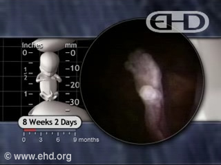
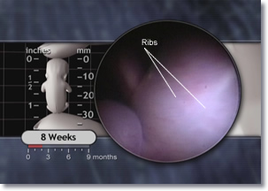
Day by day, the embryo’s bones grow. The ribs now have cartilage25 and the shafts of the long bones (in the arms and legs) begin to harden.26 The joints of the embryo already resemble adult joints in structure,27 with the elbows and the knees apparent.28 Formation of wrist ligaments begins.29

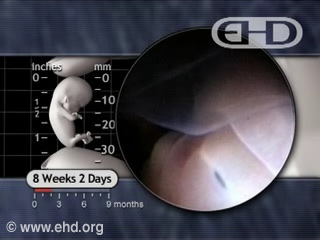
At 8 weeks, the embryo’s internal organs, before easily visible through the thin skin, become relatively hidden as the epidermis becomes a two-layered membrane.30 On the skin, eyebrows begin to appear along with fine hairs around the mouth.31

8 weeks marks the end of the embryonic period. During this time, the human embryo has grown from a single cell into nearly 1 billion cells32 forming over 4000 distinct anatomic structures. The embryo now possesses more than 90 percent of the structures found in the adult.33

