Developmental Stages in Human Embryos
Go to Stage: Intro 1 2 3 4 5 6 7 8 9 10 11 12 13 14 15 16 17 18 19 20 21 22 23
Stage 20
Page 252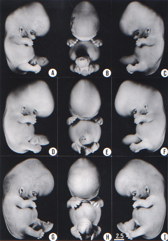
Fig. 20-1. Photographs of three embryos belonging to stage 20. A slight bend is present at the elbow, and the hands are curving medially over the cardiac region. Top row, No. 7274. Middle row. No. 7906. Bottom row. No 8157. All views are at the same magnification.
Page 253SIZE AND AGE
Most embryos of stage 20 measure 21–23 mm in length.
The age is believed to be approximately 50–51 postovulatory days.
EXTERNAL FORM
Details of internal structures may now be obscured from surface view because of the thickening mesoderm, except in fresh or formalin-fixed specimens. Thus the cerebellum isno longer distinct as seen from the surface (fig. 20-1).
The upper limbs have increased in length and become slightly bent at the elbows (fig. 20-1). The hands with their short, stubby fingers are still far apart, but they are curving slightly over the cardiac region and approach the lateral margins of the nose.
A delicate, fringe-like vascular plexus now appears in the superficial tissues of the head (fig. 20-2). In the temporofrontal region a growth center arches over the eye, and in the occipital region a second growth center occurs above the ear. The edge of the plexus is approximately halfway between the eye-ear level and the vertex of the head.
FEATURES FOR POINT SCORES
1. Cornea. The developing cornea comprises the anterior epithelium, an acellular postepithelial layer (the future substantia propria), and the posterior epithelium.
2. Optic nerve. Few nerve fibers are present. A lumen may still extend along practically the whole length of the stalk, but it is becoming obliterated (O'Rahilly, 1966, fig. 49).
3. Cochlear duct. The tip of the duct, having grown "upward," now proceeds "horizontally" (fig. 19-6).
4. Adenohypophysis. The stalk is long and slender. Capillaries are appearing in the mesoderm at the rostral surface of the adenohypophysis.
5. Vomeronasal organ. The vomeronasal organ appears as a shallow, blind sac with a broad opening (fig. 19-9).
6. Submandibular gland. The duct is longer and knobby and is situated well within the gland (fig. 19-10).
7. Metanephros. The renal vesicles are developing S-shaped lumina (fig. 19-11).
8.Humerus. The appearances are transitional between those of stages 19 and 21 (Streeter, 1949, fig. 3).
ADDITIONAL FEATURES
Blood vascular system. The arteries and veins have been illustrated by Blechschmidt (1963, plate 37).
Digestive system. A reconstruction of the pharynx and adjacent endocrine glands was published by Welter (1933, plate 1), ones of the alimentary canal by Shikinami (1926, plate 3) and by Blechschmidt (1963, plate 44).
Larynx. Three photomicrographs were published by Tucker and O'Rahilly (1972, figs. 14-16).
Urinary system. A reconstruction of the urinary system has been published by Shikinami (1926, fig. 5). Page 254 The external surface of the metanephros is said to be slightly lobulated.
Testis. From stage 20 onward the testis is said to be elliptical and have a smooth surface (Lee, 1971). It contains short, straight tubules surrounded by a darkly stained mesenchymal sheath and the rete testis. The tunica albuginea appears to be continuous with the coelomic epithelium.
Ovary. From stage 20 onward the ovary is said to be cylindrical and have a coarse surface (Lee, 1971). The presence of an ovary is first determined per exclusionem and not earlier than stage 20. Cord-like structures are absent from the cortex, and the surface epithelium shows no sign of development into a tunica albuginea. The ovary consists of "a mass of undifferentiated oogonia" (Wilson, 1926a) intermingled with fusiform mesenchymal cells and intermediate pregranulosa cells (Lee, 1971), and covered by the surface epithelium.
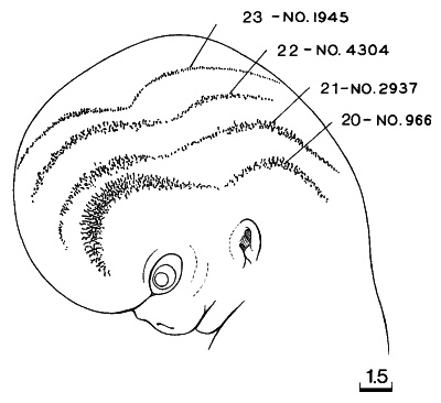
Fig. 20-2. Composite drawing showing the edge of the superficial vascular plexus in the head at stages 20-23. A subcutaneous capillary plexus that spreads upward toward the vertex of the head is a conspicuous feature of specimens measuring from about 20–24 mm in length. The transition from vascular to avascular mesoderm is more gradual in earlier stages and becomes more abrupt later.
Skeletal system. Skeletal development and the muscular system have been illustrated by Blechschmidt (1963, plates 36 and 38). The skull has been described and illustrated by Lewis (1920).
Brain. A general view of the organ can be seen in Lewis (1920, plate 3, fig. 8), and certain internal details were provided by Hines (1922, fig. 17). The nervous system as a whole was illustrated by Blechschmidt (1963, plate 35).
The hemispheres now cover two-thirds of the diencephalon. The inferior colliculus of the mesencephalon can be discerned (Bartelmez and Dekaban, 1962). Some optic fibers reach the optic chiasma. The choroid plexus of the lateral ventricles is present. The primordium of the falx cerebri begins to form (O'Rahilly and Müller, 1986b).
Eye. The lens cavity is obliterated and a lens suture begins to form.
Copyright © 1987 Carnegie Institution of Washington. Reproduced on ehd.org with permission.