Developmental Stages in Human Embryos
Go to Stage: Intro 1 2 3 4 5 6 7 8 9 10 11 12 13 14 15 16 17 18 19 20 21 22 23
Stage 22
Page 258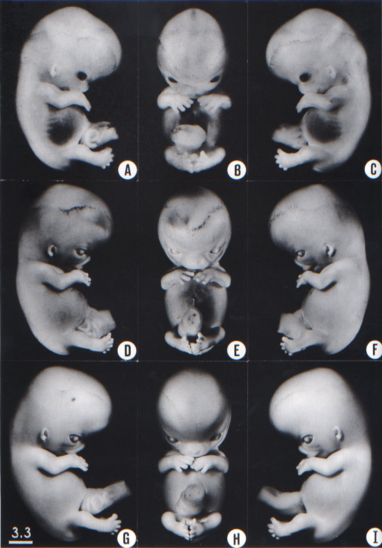
Fig. 22-1. Photographs of three embryos belonging to stage 22. The eyes are more than half covered by the eyelids. The limbs have increased in length, and the digits touch or overlap. Top row, No. 6701. Middle row, No. 6832. Bottom row, No. 8394. All views are at the same magnification.
Page 259SIZE AND AGE
The middle group of embryos of this stage measure 25–27 mm in length.
The age is believed to be approximately 54 postovulatory days.
EXTERNAL FORM
The eyelids, which have been thickening gradually, are now rapidly encroaching upon the eyes. The formation of the auricle has progressed noticeably: the tragus and antitragus especially are assuming a more definite form. The superficial vascular plexus of the head extends upward about three-quarters of the way above the eye-ear level. The hands extend further out in front of the body of the embryo, and the fingers of one hand may overlap those of the other.
FEATURES FOR POINT SCORES
1. Cornea. The cellular invasion of the postepithelial layer is complete centrally in some eyes (Streeter, 1951, fig. 17). A scleral condensation is now definite (Gilbert, 1957).
2. Optic nerve. The mesenchyme surrounding the optic nerve forms a definite sheath.
3. Cochlear duct. The tip of the duct points “upward” for the second time (fig. 19-6).
4. Adenohypophysis. Remnants of the incomplete stalk are present at each end (fig. 19-7).
5. Vomeronasal organ. The appearances are intermediate between those of stages 21 and 23 (fig. 19-9).
6. Submandibular gland. The duct shows secondary branches. It is practically solid but a suggestion of a lumen can be found in its oral part (fig. 19-10).
7. Metanephros. A few large glomeruli are present (fig. 19-12).
8. Humerus. Cartilaginous phases 1–4 are still present (Streeter, 1949, figs. 3 and 15–17). The formation of osteoblasts is beginning. A bony collar appears in the humerus, radius, ulna, and femur and tibia during stages 22 and 23 (O’Rahilly and Gardner, 1972).
ADDITIONAL FEATURES
Blood vascular system. The aortic arch system has been illustrated by Boyd (1937, fig. 1).
Heart. Reconstructions of the atrial region were reproduced by Licata (1954, figs. 4 and 5). Chordae tendineae begin to form at stages 22 and 23 (Magovern, Moore, and Hutchins, 1986).
Paramesonephric ducts. The paramesonephric ducts lie side-by-side caudally and show rostral vertical, middle transverse, and caudal vertical portions.
Brain. A general view of the organ was given by Hochstetter (1919, fig. 42).
The superior and inferior colliculi of the midbrain are indicated by their lamination (Bartelmez and Dekaban, 1962). The epithalamus is individualized by the presence of the sulcus dorsalis. In the hemispheres the cortical plate begins to appear.
Copyright © 1987 Carnegie Institution of Washington. Reproduced on ehd.org with permission.